-
|
-
The neocortex represents the evolutionary youngest part of the mammalian brain and is responsible for higher cognitive functions, sensory perception, and consciousness (Rakic, 2009). From a phylogenetic perspective, older cortical structures, including the paleocortex and archicortex, developed earlier. The paleocortex is primarily involved in olfactory processing, whereas the archicortex, which includes the hippocampus, plays a crucial role in emotional regulation, species-preserving behaviors, and memory formation (Molyneaux et al., 2007). Together, these evolutionary older regions are called allocortex and in humans they account for approximately 10% of the cerebral cortex, whereas the remaining 90% constitutes the neocortex.
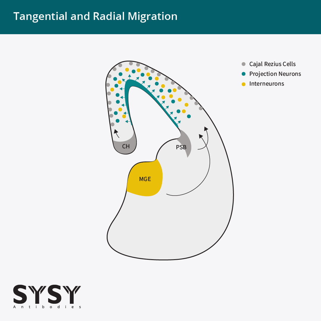
The neocortex is characterized by a highly conserved six-layered organization, with each layer defined by distinct neuronal subtypes, glial compositions, and connectivity patterns (Rakic, 2009; Molyneaux et al., 2007). Cortical neurons originate from the pallium and two proliferative zones, the ventricular zone (VZ) and subventricular zone (SVZ). Interneurons originate from subpallidal structures like the medial ganglionic eminence (MGE). Both migrate over long distances to reach their final positions within the cortex. Locally, cortical circuits are formed by interneurons, whereas long-range connections to intracortical, subcortical, and subcerebral regions are mediated by projection neurons (Rakic, 2009). The development of the neocortex is therefore a complex spatiotemporal process, characterized by radial migration of projection neurons (PN) and tangential migration of interneurons, and transient glutamatergic neurons like Cajal Retzius cells (CR cells) (Tyson & Anderson 2014; Barber & Pierani 2015, Figure 1). In this featured topic we focus on the layer markers of projection neurons during development and adulthood.
Figure 1: Tangential and radial migration.
Excitatory projection neurons show radial migration from the ventricular and subventricular zones (VZ/SVZ, green), whereas inhibitory interneurons originate from the medial ganglionic eminence (MGE, yellow) and migrate tangentially into the neocortex. Cajal Retzius cells (CR cells, gray), migrate tangentially into the marginal zone (layer I). CR cells originate from different regions, primarily from the cortical hem (CH) with additional contributions from the pallial–subpallial boundary (PSB) and septum.
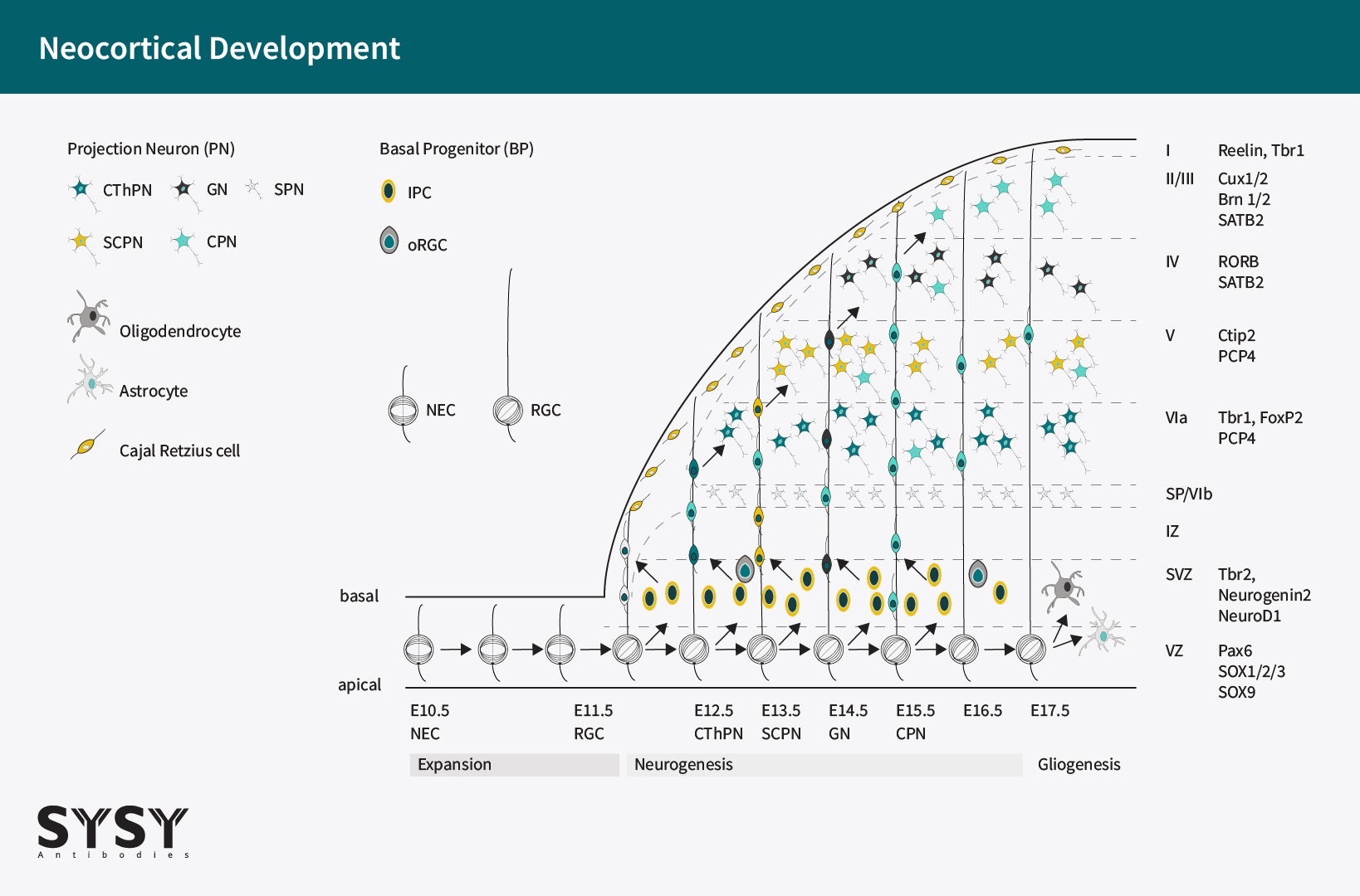
Cortical layers emerge at precisely regulated time points during early embryonic development. In mice, cortical neurogenesis begins around embryonic day 11 (E11), when progenitor cells in the ventricular zone (VZ) switch from symmetric proliferative divisions to asymmetric divisions that generate postmitotic neurons destined for radial migration (Molyneaux et al., 2007; Shitamukai and Matsuzaki, 2012). Newly born neurons migrate predominantly along radial glial fibers toward the pial surface, following an inside-out pattern of cortical layer formation. Successive waves of neurons migrate past earlier-born neurons, giving rise to progressively more superficial layers (Greig et al., 2013). Between embryonic days 16 and 18 (E16–E18), the final wave of neurons exits the ventricular zone and settles beneath the marginal zone (MZ, layer I), forming layer II and completing the six-layered cortical structure (Figure 2).
Figure 2: Illustration of neocortical development in the mouse brain.
Neuroepithelial cells (NECs) represent the earliest progenitor population of the dorsal telencephalon. They line the neural tube and undergo symmetric divisions to expand the progenitor pool, thereby establishing the founder population for all subsequent cortical lineages. Neural stem cells (NSCs), also referred to as radial glial cells (RGCs), arise from NECs and constitute the principal stem cell population of the ventricular zone (VZ). They exhibit a distinct apical–basal polarity and predominantly divide asymmetrically, generating one self-renewing progenitor and one differentiated daughter cell, thereby serving as the primary source of cortical neurons and glia. From approximately embryonic day (E) 11.5 onward, the onset of the neurogenic phase is marked by the emergence of basal progenitors (BPs). There are two types of BPs: intermediate progenitor cells (IPCs) and outer radial glial cells (oRGCs). BPs migrate into the subventricular zone (SVZ), where they lose ventricular contact and undergo mainly symmetric divisions to amplify neuronal output. IPCs and oRGCs function as transit-amplifying intermediates that markedly increase neuronal production, particularly for the upper cortical layers. IPCs are the predominant type of BP in rodents, whereas oRGCs are highly abundant in primates and have a higher amplification rate. Projection neurons are generated in a temporally ordered sequence and migrate along RGC scaffolds through the intermediate zone (IZ) to establish cortical layers in an inside-out pattern. Cajal Retzius cells (CR cells) migrate tangentially into the marginal zone (also defined as layer I), where they contribute to cortical organization. Neuronal subtype specification follows a stereotyped developmental timeline, with subplate neurons (SPNs) generated around E11.5, corticothalamic projection neurons (CThPNs) around E12.5, subcerebral projection neurons (SCPNs) around E13.5, granular neurons (GNs) around E14.5, and most callosal projection neurons (CPNs) between E14.5 and E16.5. Following neurogenesis, progenitor competence progressively shifts toward gliogenesis, giving rise to astrocytes, oligodendrocytes, and ependymal cells, while NSC potential becomes increasingly restricted. Postnatally, CR cells decline sharply through apoptosis, leaving only a sparse residual population in the adult cortex.
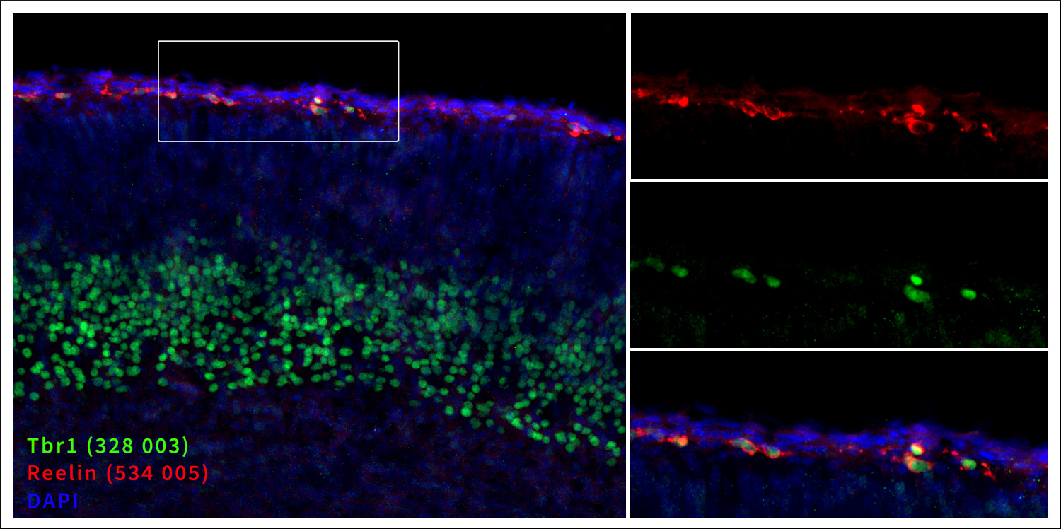
Cortical layer formation is tightly regulated by a balance of attractive and repulsive molecular cues. One of the first steps is the tangential migration of the Cajal Retzius cells (CR cells) from non-cortical regions into the marginal zone (MZ), also defined as layer I. CR cells are born between E10.5 and E13.5 (Moreau et al., 2023) and secrete Reelin, a large extracellular matrix protein and a key regulator of layer formation. Reelin promotes neuronal migration toward the cortical surface while simultaneously acting as a stop signal that instructs neurons to terminate migration at the appropriate laminar position (Richards et al., 1997; Rakic, 2009). The reeler mouse, which carries a loss-of-function mutation in the reelin gene, displays not only a "reeling gait" but also a striking inversion of cortical layering (Falconer, 1951; Caviness, 1976), underscoring the critical role of Reelin in this process. From a marker perspective, Reelin is necessary but not sufficient for identifying CR cells, as interneurons derived from the caudal ganglionic eminence (CGE) can also express Reelin (Lodato et al., 2011; Kilb and Frotscher, 2016). In this context, the expression of the transcription factor Tbr1 (T-box brain 1) serves as a useful distinguishing marker. Tbr1 is expressed in CR cells of the MZ and other glutamatergic projection neurons, but is absent in interneurons (Hevner et al., 2001; Hevner et al., 2003). Therefore, the co-expression of Reelin and Tbr1 provides a robust marker combination for identifying CR cells within the MZ (Figure 3). Furthermore, in contrast to all other neurons in the developing cerebral cortex, CR cells are FOXG1-negative (Moreau et al., 2023). Postnatally, the majority of CR cells undergo apoptosis. Consequently, the MZ of the adult brain is sparsely populated, making it easily distinguishable from the densely populated layer II.
Figure 3: Markers of the marginal zone (MZ): Reelin and Tbr1.
Indirect immunostaining of a formaldehyde-fixed mouse brain section (embryo E18, cortex) with guinea pig anti-Reelin antibody (cat. no. 534 005, dilution 1 : 500, red) and rabbit anti-Tbr1 antibody (cat. no. 328 003 dilution 1 : 500, green). Nuclei were visualized with DAPI staining (blue).
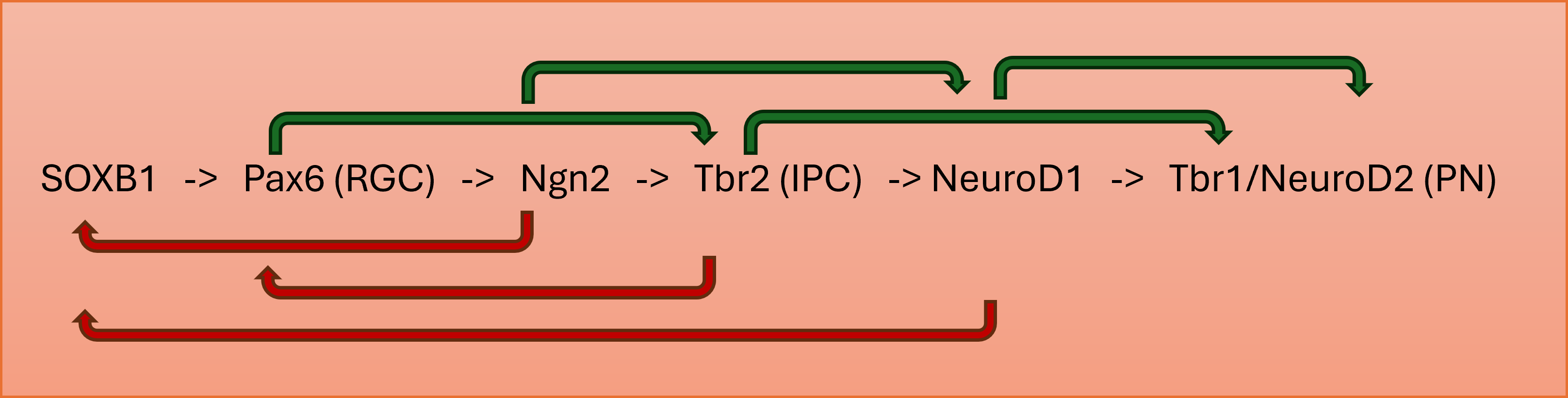
All neocortical excitatory projection neurons are generated from self-renewing progenitors of the VZ and SVZ. During the differentiation of progenitor cells into projection neurons, a sequential expression of transcription factors (TFs) has been described. This expression is linked to the differentiation state from a radial glial cell (RGC) to an intermediate progenitor cell (IPC) and finally to a projection neuron (PN) (Englund et al., 2005; Hevner, 2006; Elsen et al., 2018; Hevner, 2019; Tutucova et al., 2021, Figure 4).
Figure 4: Regulation of neural progenitor differentiation.
The differentiation of neural progenitor cells into mature neurons is regulated by a cascade of transcription factors, which includes feedforward gene activation (green arrows) and negative feedback signaling (red arrows). RGC, radial glial cell; IPC, intermediate progenitor cell; PN, projection neuron.
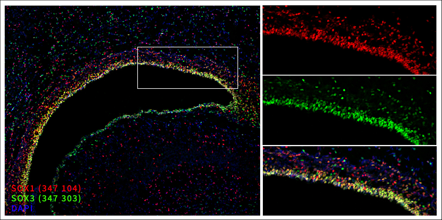
During mouse brain development, the transcription factors SOX1-3 (SRY-box transcription factors) are expressed very early during neurogenesis in the VZ by neuroepithelial cells (NECs) and, upon further differentiation by radial glial cells (RGCs) (Bylund et al., 2003; Hagey and Muhr, 2014, Figure 5). All three belong to the SOXB1 group of the SOX gene family. In contrast to SOXB1 proteins, the expression of the homeodomain transcription factor Pax6 (paired box 6) starts with the transition of NECs to RGCs; therefore, Pax6 is known as the master marker for RGCs. However, it has been debated whether Pax6 expression persists into early stages of IPCs and whether SOX9 might be a better marker for RGCs (Elsen et al., 2018). SOXB1 proteins, SOX9 and Pax6 keep neural progenitors in an undifferentiated state and suppress neuronal differentiation. Therefore, these transcription factors are important for the maintenance of stemness. In the case of neocortical development, they ensure a sufficient pool of progenitors to generate the upper layers after the deep layers are born. A suggested mechanism is that RGCs with high SOXB1 expression undergo apical symmetrical division with a slow rate (Hagey and Muhr, 2014). Reduced levels of SOXB1 proteins promote apical asymmetrical divisions, leading to one daughter cell being identical to the mother cell (self-renewal) and the other differentiating into an intermediate progenitor cell (IPC) that migrates to the subventricular zone (SVZ). Very low levels of SOXB1 proteins allow basal symmetrical divisions with increased cell cycle rates. For further differentiation into neurons, SOXB1 protein expression must be downregulated; consequently, there is almost no overlap of proneural basic helix-loop-helix (bHLH) transcription factors like NeuroD1 with SOXB1 proteins (Figure 7).
Figure 5: Markers of the ventricular zone (VZ): SOX1 and SOX3.
Indirect immunostaining of a formaldehyde-fixed rat brain section (newborn P0, ventricle) with guinea pig anti-SOX1 antibody (cat. no. 347 104, dilution 1 : 2000, red) and rabbit anti-SOX3 antibody (cat. no. 347 303, dilution 1 : 500, green). Nuclei were visualized with DAPI staining (blue).
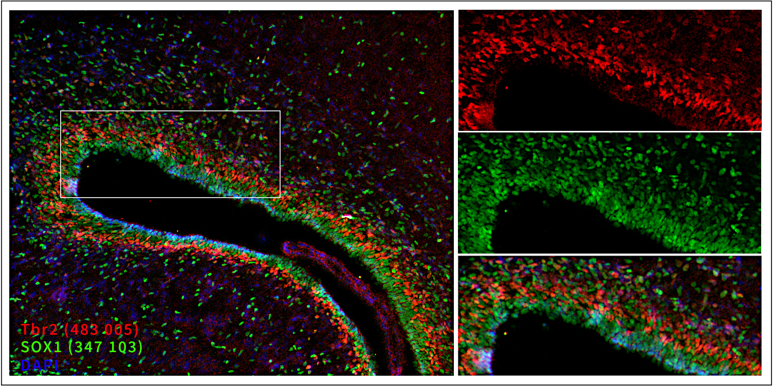
Neurogenin1 and Neurogenin2 (Ngn1 and Ngn2) are basic helix-loop-helix (bHLH) transcription factors expressed first in the VZ at E11.5–E12.5 and from E13.5 onward, in the SVZ as well. Ngn1 and Ngn2 are important for the commitment to a neuronal fate and the inhibition of glial cell fates. Therefore, Ngn1 and Ngn2 are the first markers to label a newborn cell committed to becoming a neuron. Furthermore, Ngn2 promotes further differentiation by driving the expression of NeuroD1 and downregulating the expression of the SOXB1 genes. Both are essential steps in the progression of a neural precursor into a postmitotic neuron. (Bylund et al., 2003; Evsen et al., 2013). Pax6, the marker of RGCs, activates the expression of Tbr2 (T-box brain gene 2, also known as EOMES), a T-domain transcription factor, and thereby induces its own downregulation via a negative feedback loop (Elsen et al., 2018; Hevner, 2019, Figure 4). This marks the transition from an RGC into an IPC. Tbr2 acts as the master marker for IPCs, which are cells located mainly in the SVZ (Englund et al., 2005; Hevner, 2019, Figure 6). Tbr2-positive cells still show a high proliferation rate, are important for neurogenic expansion and serve as an indicator of ongoing neurogenesis during cortical development (Englund et al., 2005). Tbr2 expression induces the activation of downstream transcription factors, such as Tbr1 and SATB2, thereby driving the differentiation of IPCs into postmitotic glutamatergic PNs.
Figure 6: Markers of the ventricular zone (VZ, SOX1) and subventricular zone (SVZ, Tbr2).
Indirect immunostaining of a formaldehyde-fixed rat brain section (embryo E18, ventricle) with rabbit anti-SOX1 antibody (cat. no. 347 103, dilution 1 : 500, green) and guinea pig anti-Tbr2 antibody (cat. no. 483 005, dilution 1 : 500, red). Nuclei were visualized with DAPI staining (blue).
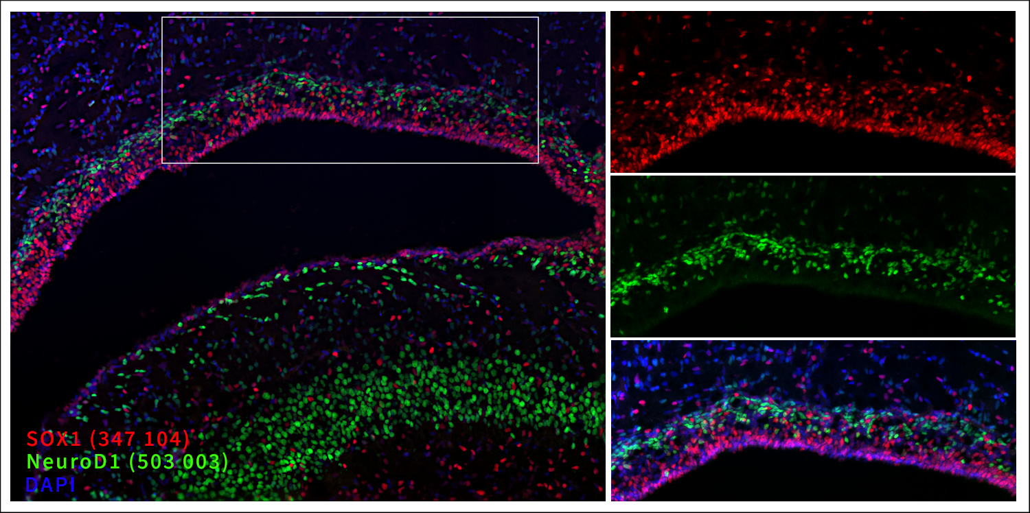
Like neurogenins, NeuroD1 belongs to the class III bHLH transcription factors and acts as a neuronal determination factor (proneural factor) that directs the cell fate toward a neuronal lineage (Bormuth, 2015; Tutucova et al., 2021). In contrast to Ngn2, NeuroD1 additionally functions as a differentiation factor, driving the mitotic progenitors to differentiate into mature postmitotic neurons. A crucial requirement for this step is the downregulation of SOXB1 genes, a process in which NeuroD1 itself is actively involved in the suppression of SOXB1 expression (Evers et al., 2013). NeuroD1 is expressed in the upper SVZ and lower intermediate zone (IZ), colocalizing within a subset of cells positive for Tbr2 or Tbr1, while showing virtually no overlap with SOXB1 proteins (Figure 7). Consequently, NeuroD1 is considered a marker for the transition from a late IPC to an early postmitotic migrating PN (Hevner, 2006). NeuroD2 acts as a differentiation factor as well and controls pyramidal neuron migration and axonal navigation (Tutucova et al., 2021).
Figure 7: Markers of the ventricular zone (VZ, SOX1) and upper subventricular zone (SVZ, NeuroD1).
Indirect immunostaining of a formaldehyde-fixed rat brain section (newborn P0, ventricle) with guinea pig anti-SOX1 antibody (cat. no. 347 104, dilution 1 : 2000, red) and rabbit anti-NeuroD1 antibody (cat. no. 503 003, dilution 1 : 2000, green).
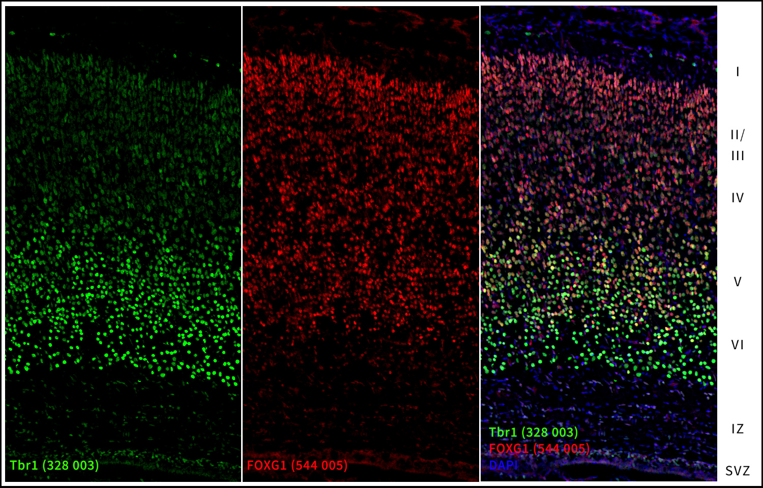
Distinct projection neuron (PN) subtypes are born in sequential waves during cortical development in an inside-out pattern (Greig et al., 2013; Figure 2). Neurogenesis begins with the generation of layer VI, followed sequentially by layers V, IV, III, and II. Later-born PNs migrate through pre-existing layers along RGCs, guided by specific signaling molecules. The first wave, peaking around E12.5, gives rise to corticothalamic projection neurons (CThPNs), which primarily reside in layer VI and express Tbr1 as a key identity gene (Figure 8). Subsequently, subcerebral projection neurons (SCPNs) are generated around E13.5, a process critically dependent on the specification factors Fezf2 and Ctip2. The next wave, at approximately E14.5, comprises granular neurons (GNs) that settle in layer IV and express the crucial marker RORB. The final wave peaks at E15.5 and produces callosal projection neurons (CPNs) destined for upper layers II/III. Notably, a subset of CPNs is born earlier and positions itself within deeper layers according to their birthdate. Established markers for CPNs include SATB2, CUX1/2, and Brn1/2. Beyond these layer-specific markers, general master regulators of brain development, such as FOXG1, play a key role in subtype specification (Hou et al., 2020; Liu et al., 2022). Mechanistically, FOXG1 directly activates SATB2 expression while differentially regulating Tbr1 and Ctip2 expression. Detailed information about CP markers is included in the next section about cortical layer markers in the adult brain.
Figure 8: Markers of the cortical plate (CP): Tbr1 and FoxG1.
Indirect immunostaining of a formaldehyde-fixed rat brain section (newborn P0, cortical plate) with rabbit anti-Tbr1 antibody (cat. no. 328 003, dilution 1 : 500, green) and guinea pig anti-FOXG1 antibody (cat. no. 544 005, dilution 1 : 1000, red). Nuclei were visualized with DAPI staining (blue).
| Marker | Reelin | SOX1-3 | Pax6 | Ngn2 | Tbr2 | NeuroD1 | Tbr1 | NeuroD2 |
| MZ, Layer 1 | ||||||||
| Layer 2 | ||||||||
| Layer 3 | ||||||||
| Layer 4 | ||||||||
| Layer 5 | ||||||||
| Layer 6 | ||||||||
| IZ/PP/SP | ||||||||
| SVZ | ||||||||
| VZ |
low medium strong
The adult neocortex contains at least 56 molecularly defined subtypes of glutamatergic PNs. During development, IPCs generate deep- and upper-layer PNs (Tasic et al., 2018; Hevner 2019; Mihalas et al., 2016). When discussing layer markers, it is crucial to understand that these proteins are expressed by distinct PN subtypes predominantly located within specific layers. Consequently, these markers are not exclusively confined to their assigned layers. Although they exhibit peak expression levels within designated layers, they may also be detectable at lower levels in adjacent regions or even show high expression in a smaller subset of cells elsewhere. Furthermore, the observed distribution pattern can be influenced by antibody affinity and the applied dilution factor, which may either emphasize layer-specific enrichment or reveal weaker expression in additional layers.
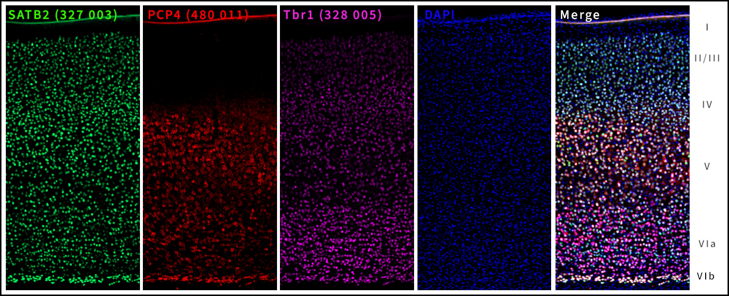
Layer VI is primarily composed of corticothalamic projection neurons (CThPN ) that send feedback connections from the cortex to the thalamus. This corticothalamic pathway is essential for modulating thalamic activity and shaping the thalamocortical information flow, thereby contributing to sensory processing and cortical state regulation. Key molecular markers of layer VI include Tbr1 (Figure 9) and FoxP2. Tbr1 is expressed early in postmitotic neurons and is required for the specification of deep-layer cortical identity, particularly influencing the differentiation and connectivity of corticothalamic projectionneurons (Hevner et al., 2001). FoxP2 is expressed in a subset of layer VI neurons and has been implicated in the regulation of thalamocortical circuitry and synaptic organization within these networks (Ferland et al., 2003). PCP4 (Purkinje cell protein 4) is used as well to identify a specific subpopulation within the deep layers, located particularly in layer VI but to some extent also in layer V (Watakabe et al., 2012). PCP4-positive PNs project to specific thalamic nuclei and PCP4 is used together with CCK (cholecystokinin) to map the sublaminar organization of the cortex.
Figure 9: Markers of deep layer VI: PCP4 and Tbr1.
Indirect immunostaining of a formaldehyde-fixed adult mouse cortex section with rabbit anti-SATB2 (cat. no. 327 003, dilution 1 : 1000, green), mouse anti-PCP4 (cat. no. 480 011, dilution 1 : 500, red) and guinea pig anti-Tbr1 (cat. no. 328 005, dilution 1 : 500, purple). Nuclei were visualized with DAPI staining (blue).
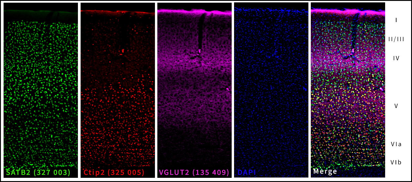
Against this molecular backdrop of cytoskeletal regulation during cortical development, it is essential to consider how these mechanisms relate to the specification and maturation of distinct neuronal populations. In particular, subcerebral projection neurons (SCPNs) of cortical layer V consist of large pyramidal neurons that project to subcortical targets such as the brainstem and spinal cord. These neurons typically express Ctip2 (Figure 10), a transcription factor essential for the development of corticospinal projections. Layer V neurons are pivotal for the execution of voluntary movements, primarily through the corticospinal tract. Ctip2 regulates axonal outgrowth, guidance, and target selection. Moreover, the loss of Ctip2 expression results in severe impairments in the formation of subcortical projections, underscoring its fundamental role in establishing the functional identity of layer V neurons (Arlotta et al., 2005; Molyneaux et al., 2007). Together, layers V and VI constitute the deep cortical layers, which play a vital role in transmitting cortical output to subcortical and thalamic structures.
Figure 10: Marker of the deep layer V: Ctip2.
Indirect immunostaining of a formaldehyde-fixed adult mouse cortex section with rabbit anti-SATB2 (cat. no. 327 003, dilution 1 : 1000, green), guinea pig anti-Ctip2 (cat. no. 325 005, dilution 1 : 500, red) and chicken anti-VGLUT2 (cat. no. 135 409, dilution 1 : 500, purple). Nuclei were visualized with DAPI staining (blue).
RORB (Retinoic Acid-Related Orphan Receptor Beta, also written as RORβ) is a highly specific transcription factor and one of the most widely used molecular markers for identifying cortical layer IV neurons in the mammalian cerebral cortex. It is predominantly expressed in thalamorecipient excitatory neurons of layer IV, where it plays an important role in neuronal differentiation, sensory circuit organization, and cortical arealization. In the murine neocortex, RORB expression forms a prominent, dense band of neurons and is particularly enriched in the barrel structures of the somatosensory cortex, which receive direct thalamic input. Beyond its strong enrichment in layer IV, lower levels of RORB expression can also be observed in subsets of neurons within layers III and V, reflecting a partial overlap with neighboring intracortical projection neuron populations. Developmentally, RORB expression emerges during cortical maturation and is closely associated with the establishment of sensory processing circuits and laminar identity. Because of its specificity and robust expression pattern, RORB is extensively used in developmental neurobiology, transcriptomic cell-type classification, and studies investigating cortical organization and connectivity.
SATB2 (Special AT-rich sequence-binding protein 2) is a key transcription factor that defines neuronal identity in the upper layers (II–IV) of the neocortex. It is strongly expressed in callosal projection neurons and is widely used as a molecular marker for this population. During cortical development, SATB2 plays a central role in neuronal differentiation, laminar specification, and the establishment of interhemispheric (callosal) connectivity. The neocortex develops in an “inside-out” pattern, where deep-layer neurons (V–VI) are generated first, followed by upper-layer neurons. SATB2 is expressed in postmitotic neurons destined for upper layers (II–IV) and establishes their identity by promoting upper-layer gene programs while actively repressing deep-layer fates, specifically by suppressing Ctip2 (BCL11B) expression. This reciprocal regulation ensures the correct molecular and functional specification of cortical projection neuron subtypes. Interestingly, beyond its prominent role in superficial layers, SATB2 also plays a specific, distinct role in the differentiation of neurons within layer 6b (the subplate). Functional studies in Satb2 knockout mice show that the loss of SATB2 leads to a fate switch: upper-layer neurons adopt deep-layer characteristics and aberrantly express corticospinal markers. As a result, callosal connectivity is strongly reduced, and neurons of the upper layers are misrouted, projecting aberrant connections to subcortical targets instead of forming proper callosal projections. Furthermore, SATB2 is important for correct cortical lamination. Without SATB2, upper-layer neurons often fail to migrate properly to superficial cortical layers, demonstrating that SATB2 is essential for the identity, positioning, and connectivity of cortical neurons during development (Alcamo et al., 2008; Britanova et al., 2008; Yang et al., 2024).
CUX1 and CUX2 (Cut Like Homeobox) are important homeobox transcription factors that regulate the late development of the cerebral cortex, particularly the formation and connectivity of neurons in the upper cortical layers (II–IV). Both proteins are strongly expressed in superficial pyramidal neurons and are often co-expressed within the same cells. Although they bind to similar DNA sequences, CUX2 generally shows faster and more transient DNA-binding activity. During cortical development, Cux genes are expressed in neural progenitor cells, with Cux2 being particularly enriched in intermediate progenitor cells. Because of this pattern, they were initially hypothesized to determine upper-layer neuronal identity. However, studies in knockout mice demonstrated that upper-layer neurons can still form, migrate, and maintain their identity in the absence of CUX1 or CUX2. Therefore, these genes are not considered master regulators of cortical layer fate. Instead, CUX2 plays a specific role in controlling the proliferation of upper-layer progenitor cells by promoting their exit from the cell cycle. This selectively regulates the number of upper-layer neurons without affecting deeper cortical layers, suggesting a role in cortical evolutionary expansion. Both Cux genes are also involved in the development of specific interneuron populations, particularly Reelin-expressing interneurons in cortical layers II–IV, where their functions are partly redundant. In mature neurons, CUX1 and CUX2 mainly regulate later stages of neuronal differentiation, influencing dendritic and axonal development to shape neuronal connectivity. Knockout studies have demonstrated that the loss of these genes leads to reduced dendritic length and branching, lower spine density, and altered spine morphology. Overall, Cux genes are essential for the proper morphology and connectivity of cortical neurons (Weiss et al., 2019).
Brn1 and Brn2 (also known as Pou3f3 and Pou3f2, respectively) belong to the POU family of homeodomain transcription factors. In the mature cortex, they are robust markers of projection neurons in cortical layers II and III, and to a lesser extent layer IV. During embryonic development, Brn1 and Brn2 regulate the temporal transition in progenitor cells from the generation of deep-layer neurons to the production of upper-layer neurons (Dominguez et al.,2013). Loss of Brn1 and Brn2, as observed in Brn1/2 double-knockout mice, results in a profound depletion of upper-layer neurons, accompanied by a dramatic reduction or complete loss of Cux1 expression (Barão et al., 2024).
The cerebral cortex fulfills complex integrative and computational functions through extensive connectivity with multiple brain regions. Cortical circuits consist of locally projecting interneurons forming short-range synaptic connections and long-range projection neurons establishing communication both within and beyond the cortex (Rakic, 2009; Molyneaux et al., 2007). Projection neurons include commissural neurons connecting the two hemispheres via the corpus callosum and corticofugal neurons projecting to subcortical targets such as the thalamus, brainstem, and spinal cord (Greig et al., 2013). During development, distinct classes of projection neurons are generated in a temporally and spatially regulated manner. In mice, callosal projection neurons mainly arise from layers II/III and layer V of the neocortex, extending axons from embryonic day (E) 16.5 onward and forming the corpus callosum over the subsequent days (Richards et al., 1997). Their specification and guidance are orchestrated by intrinsic transcriptional programs and extrinsic molecular cues, contributing to the diversity of cortical output pathways (Molyneaux et al., 2007; Greig et al., 2013). Corticothalamic and corticospinal neurons establish bidirectional communication between the cortex and subcortical structures. Thalamocortical connectivity is particularly critical, as thalamic afferents provide major sensory input and support the functional maturation of cortical layers. These projections are densely concentrated in layer IV and, to a lesser extent, in layer VI, marking these layers as primary recipients of subcortical input (Jones, 2001). Cortical layers have traditionally been defined by layer-specific transcription factors and nuclear-associated proteins. However, synaptic proteins and axonally transported markers provide complementary insights into cortical organization. Incoming axons, particularly from the thalamus, introduce proteins that are not endogenously expressed by cortical neurons, thereby serving as indirect markers of cortical connectivity (Kaneko and Fujiyama, 2002; Fremeau et al., 2004). A prominent example is the vesicular glutamate transporter 2 (VGLUT2) (Figure 11), which is highly expressed in thalamic relay neurons but largely absent from intrinsic cortical pyramidal neurons. VGLUT2 immunoreactivity in the cortex therefore reflects thalamocortical terminals rather than cortical glutamatergic neurons, which primarily express VGLUT1. VGLUT2-positive terminals are enriched and most dense in layer IV and, to a lesser extent, in layer VI, mirroring the laminar distribution of thalamic inputs (Nahmani and Erisir, 2005; El Mestikawy et al., 2011). For further details see also “VGLUT as Marker for Glutamatergic Neurons”.
Figure 11: Projection neuron terminals as cortical layer marker: VGLUT2.
Direct immunostaining of a formaldehyde-fixed mouse brain section with Sulfo-Cyanine 3 conjugated mouse anti-VGLUT2 (cat. no. 135 421C3, dilution 1 : 500, red) and Sulfo-Cyanine 5 conjugated mouse anti-VGLUT1 (cat. no. 135 011C5, dilution 1 : 500, green). Nuclei were visualized with DAPI staining (blue).
Together, the combined use of transcriptional, nuclear and synaptic markers enables a comprehensive understanding of cortical development, laminar organization, and input–output circuitry, providing a foundation for studies of cortical function and plasticity.
Accordingly, the following heatmap illustrates the regions of highest relative protein expression while also indicating layers in which the proteins remain detectable at lower intensities.
| Marker | Tbr1 | FoxP2 | PCP4 | Ctip2 | RORB | VGLUT2 | SATB2 | Brn1/2 | CUX1/2 |
| Layer 1 | |||||||||
| Layer 2 | |||||||||
| Layer 3 | |||||||||
| Layer 4 | |||||||||
| Layer 5 | |||||||||
| Layer 6 |
low medium strong
| Cat. No. | Product Description | Application | Quantity | Price | Cart |
|---|
| 135 416 | VGLUT2, chicken, polyclonal, affinity purifiedaffinity | WB ICC IHC IHC-P (FFPE) | 50 µg | $390.00 |  |
| 135 418 | VGLUT2, Guinea pig, monoclonal, recombinant IgGrecombinant IgG K.O. | WB ICC IHC IHC-P (FFPE) ExM | 50 µg | $420.00 |   |
| 135 421 | VGLUT2, mouse, monoclonal, purified IgG IgG K.O. | WB IP ICC IHC IHC-P (FFPE) IHC-G ExM | 100 µg | $425.00 | |
| 135 421AbOR | VGLUT2, mouse, monoclonal, purified IgG IgG, AbberiorStar ORANGE | ICC | 100 µg | $470.00 | |
| 135 421AbRED | VGLUT2, mouse, monoclonal, purified IgG IgG, AbberiorStar RED | ICC | 100 µg | $470.00 | |
| 135 421BT | VGLUT2, mouse, monoclonal, purified IgG IgG, biotin | WB IHC IHC-P (FFPE) | 100 µg | $470.00 | |
| 135 421C3 | VGLUT2, mouse, monoclonal, purified IgG IgG, Sulfo-Cyanine 3 | ICC IHC | 100 µg | $475.00 | |
| 135 421C5 | VGLUT2, mouse, monoclonal, purified IgG IgG, Sulfo-Cyanine 5 | IHC | 100 µg | $470.00 | |
| 135-4P | VGLUT2, control proteincontrol protein | 100 µg | $110.00 | |
Authors: Dr. Christian Erck and Dr. Liane Wüstefeld
Christian Erck, CEO of Synaptic Systems GmbH, has a background in mouse knockout research and antibody development. Having generated his first monoclonal antibody in 1993, he now leads the monoclonal antibody department in Braunschweig.
Liane Wüstefeld heads the histology department, focusing primarily on neuroscience. As an expert in histology, she validates our in-house developed antibodies for immunohistochemistry (IHC) applications.


Arlotta et al., 2005: Neuronal subtype-specific genes that control corticospinal motor neuron development in vivo. PMID: 15664173
Alcamo et al., 2008: Satb2 Regulates Callosal Projection Neuron Identity in the Developing Cerebral Cortex. PMID: 18255030
Barão et al., 2024: BRN1/2 Function in Neocortical Size Determination and Microcephaly. PMID: 37961182
Barber and Pierani, 2015: Tangential migration of glutamatergic neurons and cortical patterning during development: Lessons from Cajal-Retzius cells. PMID: 26581033
Bormuth, 2015: Roles of bHLH Transcription Factors Neurod1, Neurod2 and Neurod6 in Cerebral Cortex Development and Commissure Formation. Dissertation.
Britanova et al., 2008: Satb2 Is a Postmitotic Determinant for Upper-Layer Neuron Specification in the Neocortex. PMID: 18255031
Bylund et al., 2003: Vertebrate neurogenesis is counteracted by Sox1-3 activity. PMID: 14517545
Caviness, 1976: Patterns of Cell and Fiber Distribution in the Neocortex of the Reeler Mutant Mouse. PMID: 1002868
Dominguez et al., 2013: POU-III transcription factors (Brn1, Brn2, and Oct6) influence neurogenesis, molecular identity, and migratory destination of upper-layer cells of the cerebral cortex. PMID: 22892427
Elsen et al., 2018: The Epigenetic Factor Landscape of Developing Neocortex Is Regulated by Transcription Factors Pax6→ Tbr2→ Tbr1. PMID: 30186101
Englund et al., 2005: Pax6, Tbr2, and Tbr1 are expressed sequentially by radial glia, intermediate progenitor cells, and postmitotic neurons in developing neocortex. PMID: 15634788
El Mestikawy et al., 2011: From glutamate co-release to vesicular synergy: vesicular glutamate transporters. PMID: 21415847
Evsen et al., 2013: Progression of neurogenesis in the inner ear requires inhibition of Sox2 transcription by neurogenin1 and neurod1. PMID: 23447599
Falconer, 1951: Two new mutants, 'trembler' and 'reeler', with neurological actions in the house mouse (Mus musculus L.). PMID: 24539699
Ferland et al., 2003: Characterization of Foxp2 and Foxp1 mRNA and protein in the developing and mature brain. PMID: 12687690
Fremeau et al., 2004: Vesicular glutamate transporters 1 and 2 target to functionally distinct synaptic release sites. PMID: 15118123
Greig et al., 2013: Molecular logic of neocortical projection neuron specification, development, and diversity. PMID: 24105342
Hagey and Muhr, 2014: Sox2 acts in a dose-dependent fashion to regulate proliferation of cortical progenitors. PMID: 25482558
Hevner et al., 2001: Tbr1 regulates differentiation of the preplate and layer 6. PMID: 11239428
Hevner et al., 2003: Cajal–Retzius cells in the mouse: transcription factors, neurotransmitters, and birthdays suggest a pallial origin. PMID: 12644247
Hevner et al., 2002: Cortical and thalamic axon pathfinding defects in Tbr1, Gbx2, and Pax6 mutant mice: evidence that cortical and thalamic axons interact and guide each other. PMID: 11967891
Hevner, 2006: From radial glia to pyramidal-projection neuron: transcription factor cascades in cerebral cortex development. PMID: 16388109
Hevner, 2019: Intermediate progenitors and Tbr2 in cortical development. PMID: 30677129
Hirota and Nakajima, 2020: VLDLR is not essential for reelin-induced neuronal aggregation but suppresses neuronal invasion into the marginal zone. PMID: 32540847
Hosseini et al., 2022: The role of α-tubulin tyrosination in controlling the structure and function of hippocampal neurons. PMID: 36340693
Hou et al., 2020: Transcription and Beyond: Delineating FOXG1 Function in Cortical Development and Disorders. PMID: 32158381
Jones, 2001: The thalamic matrix and thalamocortical synchrony. Trends Neurosci 24:595–601. PMID: 11576674
Kaneko and Fujiyama, 2002: Complementary distribution of vesicular glutamate transporters in the central nervous system. PMID: 11985876
Kilb and Frotscher, 2016: Cajal-Retzius-Zellen: Architekten der kortikalen Entwicklung. DOI10.1007/s12269-016-0058-0
Liu et al., 2022: FOXG1 sequentially orchestrates subtype specification of postmitotic cortical projection neurons. PMID: 35613274
Lodato et al., 2011: Excitatory projection neuron subtypes control the distribution of local inhibitory interneurons in the cerebral cortex. PMID: 21338885
Mihalas et al., 2016: Intermediate Progenitor Cohorts Differentially Generate Cortical Layers and Require Tbr2 for Timely Acquisition of Neuronal Subtype Identity. PMID: 27320921
Mukhtar and Taylor, 2018: Untangling Cortical Complexity During Development. PMID: 29551911
Molyneaux et al., 2007: Neuronal subtype specification in the cerebral cortex. PMID: 17514196
Moreau et al., 2023: Repurposing of the multiciliation gene regulatory network in fate specification of Cajal-Retzius neurons. PMID: 37321213
Nahmani and Erisir, 2005: VGluT2 immunochemistry identifies thalamocortical terminals in layer 4 of adult and developing visual cortex. PMID: 15770654
Rakic, 2009: Evolution of the neocortex: a perspective from developmental biology. PMID: 19763105
Richards et al., 1997: Directed growth of early cortical axons is influenced by a chemoattractant released from an intermediate target. PMID: 9065505
Shitamukai and Matsuzaki, 2012: Control of asymmetric cell division of mammalian neural progenitors. PMID: 22524601
Tasic et al., 2018: Shared and distinct transcriptomic cell types across neocortical areas. PMID: 30382198
Tutucova et al., 2021: The Role of Neurod Genes in Brain Development, Function, and Disease. PMID: 34177462
Tyson and Anderson, 2014: GABAergic interneuron transplants to study development and treat disease. PMID: 24508416
Watakabe et al., 2012: Area-specific substratification of deep layer neurons in the rat cortex. PMID: 22678985
Weiss et al., 2019: The crux of Cux genes in neuronal function and plasticity. PMID: 29518378
Yang et al., 2024: Spatial transcriptome reveals the region-specific genes and pathways regulated by Satb2 in neocortical development. PMID: 39095712
Certificates
ISO 9001 2015 Quality Management System and Green Lab Platinum certification level for sustaining laboratory processes.


Newsletter
Sign up for our newsletter and get the latest updates and news.