
-
|
-
In this step the proteins are transferred from the gel to a blotting membrane driven by an electric field. Nitrocellulose and PVDF (polyvinylidene fluoride) membranes are the most common membrane types for Western blotting.
Nitrocellulose membranes are made from nitrated cellulose and bind proteins mainly through hydrophobic interactions and hydrogen bonding.
PVDF is a hydrophobic, synthetic polymer membrane with very high protein-binding capacity. It must be pre-wetted in methanol before use.
Both membrane types are available in two typical pore sizes:
Semi-Dry blotting: This is a very economic method (time and consumables) and widely used standard approach that can be used for a broad range of proteins. In a horizontal setup the gel–membrane sandwich is placed between buffer-soaked filter papers and sandwiched between plate electrodes. It requires less buffer and is faster than wet transfer. However, it may be less efficient for very large proteins and can be more sensitive to drying artifacts if not carefully controlled.
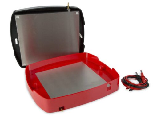
Figure 1: Semi-dry Western blot device
Figure 1: Semi-dry Western blot device
Wet (Tank)-blotting: The gel and membrane are assembled in a transfer sandwich submerged in transfer buffer within a tank. An electric field drives proteins from the gel onto the membrane. This method is highly efficient and reliable, particularly for large proteins, but it is time-consuming and requires relatively large buffer volumes.
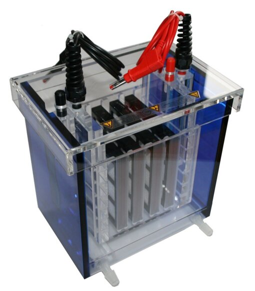
Figure 2: Wet (Tank)-blot device
Figure 2: Wet (Tank)-blot device
In general, transfer parameters are optimized for each blotting device. It is important to follow the manufacturer's instructions.
The transfer efficiency can be evaluated by using reversible staining solutions like Ponceau red or commercial products compatible with the blotting membrane.
Certificates
ISO 9001 2015 Quality Management System and Green Lab Platinum certification level for sustaining laboratory processes.


Newsletter
Sign up for our newsletter and get the latest updates and news.