Cat. No.: 250 204
Amount: 100 µl
Price:
$370.00
|
|

|
|
| Cat. No. 250 204 |
100 µl antiserum, lyophilized. For reconstitution add 100 µl H2O, then aliquot and store at -20°C until use. Antibodies should be stored at +4°C when still lyophilized. Do not freeze! |
| Applications | |
| Immunogen | Synthetic peptide corresponding to AA 146 to 161 from mouse EAAT2 (UniProt Id: P43006) |
| Reactivity |
Reacts with: rat (P31596), mouse (P43006). Other species not tested yet. Predicted to cross-react with human (P43004) due to high sequence homology. |
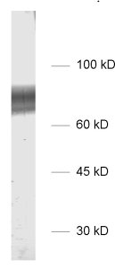
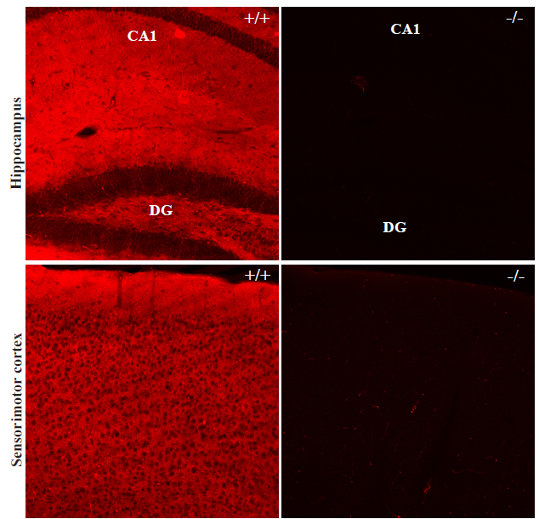
| Specificity | K.O. validated |
| Matching control protein/peptide | 250-2P |
| Data sheet | Datasheet 250_204 |
|
|
Glutamate is the major excitatory neurotransmitter in the mammalian central nervous system. After the release of glutamate from synaptic vesicles into the synaptic cleft during neurotransmission, excitatory amino acid transporters (EAATs) remove extracellular glutamate to avoid excitotoxic levels (1).
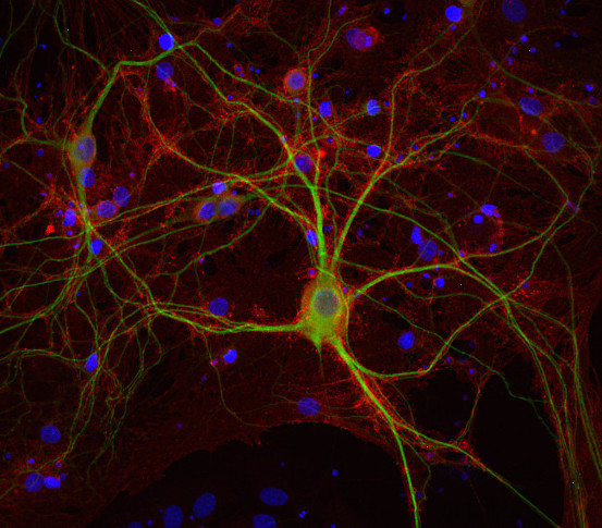
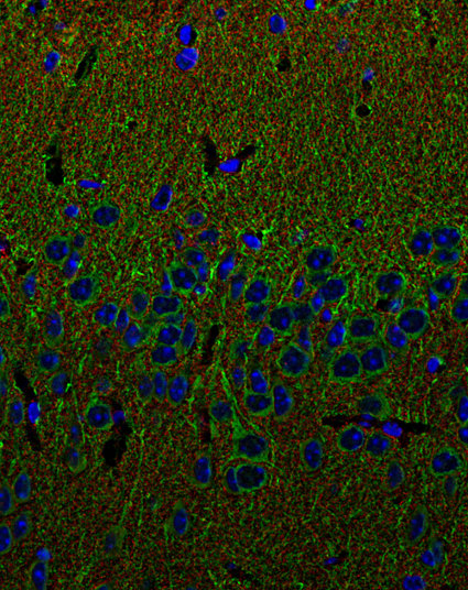
Five EAATs with differential expression patterns have been described so far: EAAT1, also referred to as GLAST and SLC1A3, has neuroprotective potential following ischemia and occurs in reactive astrocytes and activated microglia. EAAT2 (GLT-1, SLC1A2) is the most abundant isoform and is primarily expressed in astrocytes. Both variants show high levels in brain and retina. EAAT3 / SLC1A1, EAAT4 / SLC1A6 and EAAT5 / SLC1A7 are expressed in neurons (2). EAAT4 shows weak expression in the forebrain and high levels in the cerebellum, where it mainly locates to Purkinje cells (3). EAAT5 primarily occurs in the retina, where it locates very close to glutamate release sites. In K.O. mice flicker resolution is considerably compromised (4). Recent findings suggest that EAAT5 is an abundant isoform, expressed also in non-neuronal peripheral tissues (5).
Certificates
ISO 9001 2015 Quality Management System and Green Lab Platinum certification level for sustaining laboratory processes.


Newsletter
Sign up for our newsletter and get the latest updates and news.