Targeting lysosomal acidification to restore microglial homeostasis and mitigate memory decline during male brain ageing.Barrella L, Ramírez-Ponce MP, Vázquez-Román V, Millán-Huang MS, Maldonado MD, Flores-Cordero JA, Alés E
Brain, behavior, and immunity (2025) 131: 106170.
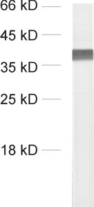
101 002 WB; tested species: mouse
Vesicular Glutamate Transporter 3 Is Involved in Glutamatergic Signalling in Podocytes.Nishii N, Kawai T, Yasuoka H, Abe T, Tatsumi N, Harada Y, Miyaji T, Li S, Tsukano M, Watanabe M, Ogawa D, et al.
International journal of molecular sciences (2025) 266: .
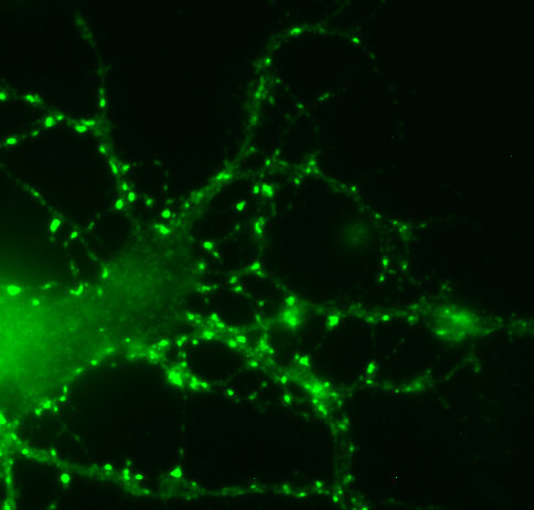
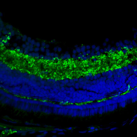
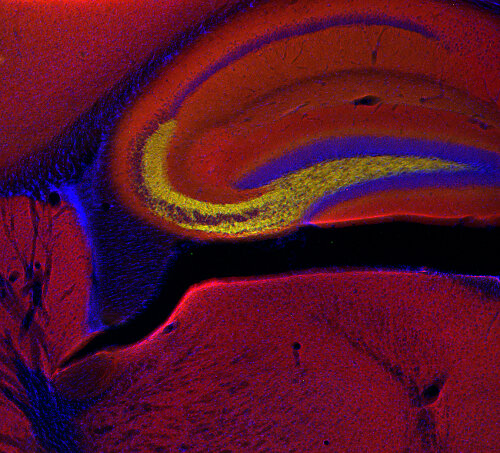
101 002 WB, ICC, IHC; tested species: mouse,rat
Impaired Response to Mismatch Novelty in the Li2+-Pilocarpine Rat Model of TLE: Correlation with Hippocampal Monoaminergic Inputs.Nascimento C, Guerreiro-Pinto V, Pawlak S, Caulino-Rocha A, Amat-Garcia L, Cunha-Reis D
Biomedicines (2024) 123: .
101 002 WB; tested species: rat
Genetically modified E. Coli secreting melanin (E.melanin) activates the astrocytic PSAP-GPR37L1 pathway and mitigates the pathogenesis of Parkinson's disease.Kong W, Liu Y, Ai P, Bi Y, Wei C, Guo X, Cai Z, Gao G, Hu P, Zheng J, Liu J, et al.
Journal of nanobiotechnology (2024) 221: 690.
101 002 WB, IHC; tested species: mouse
The CB1 receptor interacts with cereblon and drives cereblon deficiency-associated memory shortfalls.Costas-Insua C, Hermoso-López A, Moreno E, Montero-Fernández C, Álvaro-Blázquez A, Maroto IB, Sánchez-Ruiz A, Diez-Alarcia R, Blázquez C, Morales P, Canela EI, et al.
EMBO molecular medicine (2024) : .
101 002 WB; tested species: mouse
Presynapses contain distinct actin nanostructures.Bingham D, Jakobs CE, Wernert F, Boroni-Rueda F, Jullien N, Schentarra EM, Friedl K, Da Costa Moura J, van Bommel DM, Caillol G, Ogawa Y, et al.
The Journal of cell biology (2023) 22210: .
101 002 WB; tested species: rat
S-acylation of the Wnt receptor Frizzled-5 by zDHHC5 controls its cellular localization and synaptogenic activity in the rodent hippocampus.Teo S, Bossio A, Stamatakou E, Pascual-Vargas P, Jones ME, Schuhmacher LN, Salinas PC
Developmental cell (2023) 5820: 2063-2079.e9.
101 002 WB; tested species: rat
Glutamatergic transmission and receptor expression in the synucleinopathy h-α-synL62 mouse model: Effects of hydromethylthionine.Schwab K, Chasapopoulou Z, Frahm S, Magbagbeolu M, Cranston A, Harrington CR, Wischik CM, Theuring F, Riedel G
Cellular signalling (2022) 97: 110386.
101 002 WB; tested species: mouse
Colocalization of different neurotransmitter transporters on synaptic vesicles is sparse except for VGLUT1 and ZnT3.Upmanyu N, Jin J, Emde HV, Ganzella M, Bösche L, Malviya VN, Zhuleku E, Politi AZ, Ninov M, Silbern I, Leutenegger M, et al.
Neuron (2022) : .
101 002 WB, EM, UPTAKE; tested species: rat
Control of synaptic vesicle release probability via VAMP4 targeting to endolysosomes.Ivanova D, Dobson KL, Gajbhiye A, Davenport EC, Hacker D, Ultanir SK, Trost M, Cousin MA
Science advances (2021) 718: .
101 002 WB; tested species: mouse
The phosphoprotein Synapsin Ia regulates the kinetics of dense-core vesicle release.Yang HJ, Chen PC, Huang CT, Cheng TL, Hsu SP, Chen CY, Lu JC, Wang CT
The Journal of neuroscience : the official journal of the Society for Neuroscience (2021) : .
101 002 WB, ICC
Chondroitin sulfate proteoglycan-5 forms perisynaptic matrix assemblies in the adult rat cortex.Pintér A, Hevesi Z, Zahola P, Alpár A, Hanics J
Cellular signalling (2020) 74: 109710.
101 002 WB; tested species: rat
Auxiliary α2δ1 and α2δ3 Subunits of Calcium Channels Drive Excitatory and Inhibitory Neuronal Network Development.Bikbaev A, Ciuraszkiewicz-Wojciech A, Heck J, Klatt O, Freund R, Mitlöhner J, Enrile Lacalle S, Sun M, Repetto D, Frischknecht R, Ablinger C, et al.
The Journal of neuroscience : the official journal of the Society for Neuroscience (2020) 4025: 4824-4841.
101 002 WB; tested species: mouse
Rapid purification and metabolomic profiling of synaptic vesicles from mammalian brain.Chantranupong L, Saulnier JL, Wang W, Jones DR, Pacold ME, Sabatini BL
eLife (2020) 9: .
101 002 WB; tested species: mouse
Phospholipase D1 Ablation Disrupts Mouse Longitudinal Hippocampal Axis Organization and Functioning.Santa-Marinha L, Castanho I, Silva RR, Bravo FV, Miranda AM, Meira T, Morais-Ribeiro R, Marques F, Xu Y, Point du Jour K, Wenk M, et al.
Cell reports (2020) 3012: 4197-4208.e6.
101 002 WB; tested species: mouse
Endophilin-A coordinates priming and fusion of neurosecretory vesicles via intersectin.Gowrisankaran S, Houy S, Del Castillo JGP, Steubler V, Gelker M, Kroll J, Pinheiro PS, Schwitters D, Halbsgut N, Pechstein A, van Weering JRT, et al.
Nature communications (2020) 111: 1266.
101 002 WB; tested species: mouse
Uncoupling endosomal CLC chloride/proton exchange causes severe neurodegeneration.Weinert S, Gimber N, Deuschel D, Stuhlmann T, Puchkov D, Farsi Z, Ludwig CF, Novarino G, López-Cayuqueo KI, Planells-Cases R, Jentsch TJ, et al.
The EMBO journal (2020) : e103358.
101 002 WB, IHC; tested species: mouse
Monitoring translation in synaptic fractions using a ribosome profiling strategy.Simbriger K, Amorim IS, Chalkiadaki K, Lach G, Jafarnejad SM, Khoutorsky A, Gkogkas CG
Journal of neuroscience methods (2019) : 108456.
101 002 WB; tested species: mouse
Cyclophillin A deficiency accelerates RML-induced prion disease.Bouybayoune I, Comerio L, Pasetto L, Bertani I, Bonetto V, Chiesa R
Neurobiology of disease (2019) : 104498.
101 002 WB; tested species: mouse
LRRK2 modifies α-syn pathology and spread in mouse models and human neurons.Bieri G, Brahic M, Bousset L, Couthouis J, Kramer NJ, Ma R, Nakayama L, Monbureau M, Defensor E, Schüle B, Shamloo M, et al.
Acta neuropathologica (2019) : .
101 002 WB; tested species: mouse
Altered TAOK2 activity causes autism-related neurodevelopmental and cognitive abnormalities through RhoA signaling.Richter M, Murtaza N, Scharrenberg R, White SH, Johanns O, Walker S, Yuen RKC, Schwanke B, Bedürftig B, Henis M, Scharf S, et al.
Molecular psychiatry (2018) : .
101 002 WB; tested species: mouse
Isolation of Synaptic Vesicles from Genetically Engineered Cultured Neurons.McKenzie C, Spanova M, Johnson A, Kainrath S, Zheden V, Sitte HH, Janovjak H
Journal of neuroscience methods (2018) : .
101 002 WB; tested species: rat
Hypothalamic CNTF volume transmission shapes cortical noradrenergic excitability upon acute stress.Alpár A, Zahola P, Hanics J, Hevesi Z, Korchynska S, Benevento M, Pifl C, Zachar G, Perugini J, Severi I, Leitgeb P, et al.
The EMBO journal (2018) : .
101 002 WB; tested species: mouse
Regulation of the Hippocampal Network by VGLUT3-Positive CCK- GABAergic Basket Cells.Fasano C, Rocchetti J, Pietrajtis K, Zander JF, Manseau F, Sakae DY, Marcus-Sells M, Ramet L, Morel LJ, Carrel D, Dumas S, et al.
Frontiers in cellular neuroscience (2017) 11: 140.
101 002 WB, IP; tested species: mouse
PRRT2 Is a Key Component of the Ca(2+)-Dependent Neurotransmitter Release Machinery.Valente P, Castroflorio E, Rossi P, Fadda M, Sterlini B, Cervigni RI, Prestigio C, Giovedì S, Onofri F, Mura E, Guarnieri FC, et al.
Cell reports (2016) 151: 117-31.
101 002 WB
Synaptophysin 1 Clears Synaptobrevin 2 from the Presynaptic Active Zone to Prevent Short-Term Depression.Rajappa R, Gauthier-Kemper A, Böning D, Hüve J, Klingauf J
Cell reports (2016) 146: 1369-1381.
101 002 WB, ICC; tested species: rat
The GTPase Rab26 links synaptic vesicles to the autophagy pathway.Binotti B, Pavlos NJ, Riedel D, Wenzel D, Vorbrüggen G, Schalk AM, Kühnel K, Boyken J, Erck C, Martens H, Chua JJ, et al.
eLife (2015) 4: e05597.
101 002 WB, ICC
Differential spatial expression and subcellular localization of CtBP family members in rodent brain.Hübler D, Rankovic M, Richter K, Lazarevic V, Altrock WD, Fischer KD, Gundelfinger ED, Fejtova A
PloS one (2012) 76: e39710.
101 002 WB; tested species: mouse
N-cofilin can compensate for the loss of ADF in excitatory synapses.Görlich A, Wolf M, Zimmermann AM, Gurniak CB, Al Banchaabouchi M, Sassoè-Pognetto M, Witke W, Friauf E, Rust MB
PloS one (2011) 610: e26789.
101 002 WB; tested species: mouse
Lack of synapsin I reduces the readily releasable pool of synaptic vesicles at central inhibitory synapses.Baldelli P, Fassio A, Valtorta F, Benfenati F
The Journal of neuroscience : the official journal of the Society for Neuroscience (2007) 2749: 13520-31.
101 002 WB; tested species: mouse