Cat. No.: N3802-At488-L
Amount: 200 µl
Price:
$540.00
|
|

This product was developed by |
Camelid single domain antibodies (sdAbs) consist only of one antigen binding site of an Alpaca heavy chain antibody. With only ~15 kDa, these Tags are about 10-times smaller than conventional IgG antibody molecules.
|
|
| Cat. No. N3802-At488-L |
200 µl purified antibody, lyophilized from PBS, fluorescence-labeled with
ATTO® 488.
Albumin was added for stabilization. For reconstitution refer to the NanoTag reconstitution and storage instructions. Reconstitute immediately upon receipt! Avoid bright light when working with the antibody to minimize photo bleeching of the fluorescent dye. |
| Applications | |
| Label | ATTO 488, two fluorophores coupled to one FluoTag |
| Clone | 1D12 |
| Subtype | single domain |
| Immunogen | Recombinant protein corresponding to AA 1 to 432 from human GFAP (UniProt Id: P14136) |
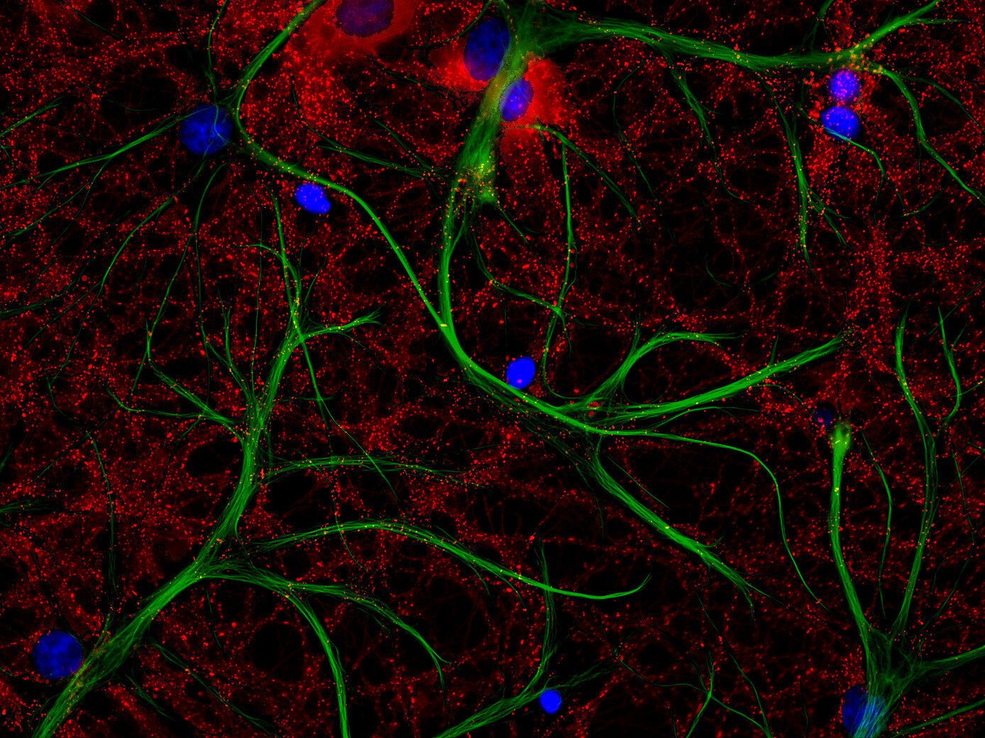
| Reactivity |
Reacts with: human (P14136), rat (P47819), mouse (P03995). Other species not tested yet. |
| Specificity | Specific for GFAP, detects all isoforms. |
| Matching control protein/peptide | 173-0P |
|
|
Glial fibrillary acidic protein GFAP is a glial-specific member of the intermediate filament protein family. This group comprises celltype-specific filamentous proteins with similar structure and function as scaffold for cytoskeleton assembly and maintenance.
Frequently, neural stem cells also express GFAP. In addition many types of brain tumors, probably derived from astrocytic cells, heavily express GFAP. This protein is also found in the lens epithelium, Kupffer cells of the liver, in some cells in salivary tumors and others.
Point-mutations in the GFAP gene have been correlated to Alexander disease a fatal leukoencephalopathy that leads to the dysmyelination or demyelination of the central nervous system.
Unlabeled variants and several modifications of sdAbs like biotin, fluorophore or DBCO conjugation are available.
In FluoTag®-X2 two fluorophore molecules are site-specifically coupled to each FluoTag molecule. Therefore, the reagent simultaneously targets two fluorophores to the protein of interest, which ensures up to two-fold („2X“)-brighter signals. Owing to the small size of the FluoTags, the distance between the target epitope and each fluorophore is ~ 3 nm.
In comparison to detection systems using conventional antibodies, FluoTag-X can thus improve the localization accuracy by 10-15 nm. Both features - superior brightness and precise fluorophore placement - render the FluoTag-X products excellent tools for all microscopy techniques.
Certificates
ISO 9001 2015 Quality Management System and Green Lab Platinum certification level for sustaining laboratory processes.


Newsletter
Sign up for our newsletter and get the latest updates and news.