Cat. No.: N0304-At488-L
Amount: 200 µl
Price:
$540.00
|
|

This product was developed by |
Camelid single domain antibodies (sdAbs) consist only of one antigen binding site of an Alpaca heavy chain antibody. With only ~15 kDa, these Tags are about 10-times smaller than conventional IgG antibody molecules.
|
|
| Cat. No. N0304-At488-L |
200 µl purified antibody, lyophilized from PBS, fluorescence-labeled with
ATTO® 488.
Albumin was added for stabilization. For reconstitution refer to the NanoTag reconstitution and storage instructions. Reconstitute immediately upon receipt! Avoid bright light when working with the antibody to minimize photo bleeching of the fluorescent dye. |
| Applications | |
| Label | ATTO 488, two fluorophores coupled to two FluoTags each |
| Clone | 1H1-1B2 |
| Subtype | single domain |
| Immunogen | Recombinant protein corresponding to AA 1 to 238 from jellyfish GFP (UniProt Id: P42212) |
| Specificity |
Recognizes GFP, mEGFP, superfolder GFP, most common CFP and YFP variants. Does not cross-react with mCherry, mRFP, dsRed, mTagBFP or their most common derivatives. |
|
|
Green fluorescent protein GFP and its derivates have become very popular and universal tools in cell biology. It is a monomeric and fast maturating protein with high photostability. Due to its sensitivity to pH changes it can be used as a biological pH indicator.
Unlabeled variants and several modifications of sdAbs like biotin, fluorophore or DBCO conjugation are available.
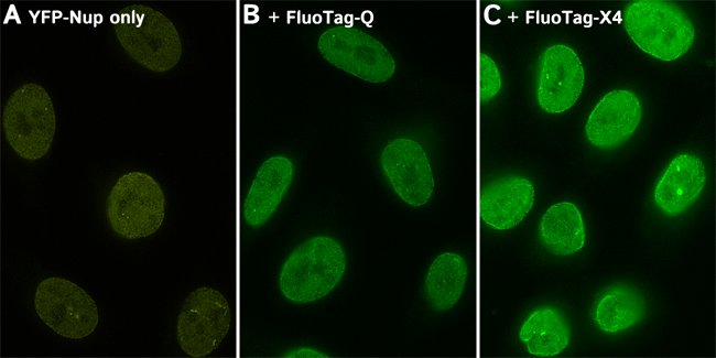
In FluoTag®-Q each fluorophore is coupled to exactly one FluoTag, which in turn binds to its target molecule in a monovalent fashion. The high binding affinity and a coupling efficiency of > 95% guarantees a highly linear relation between the number of target molecules and the intensity of fluorescence. This enables a direct count of the target molecule of interest. The fluorophore is located exceptionally close to the recognized epitope (< 1.5 nm), which is ideal for all microscopy techniques.
In FluoTag®-X two fluorophore molecules are site-specifically coupled to each FluoTag molecule. Therefore, the reagent simultaneously targets up to four fluorophores (in X4 variants) to the protein of interest, which ensures extra-bright signals. Owing to the small size of the FluoTags, the distance between the target epitope and each fluorophore is ~ 3 nm.
In comparison to detection systems using conventional antibodies, FluoTag-X can thus improve the localization accuracy by 10-15 nm. Both features - superior brightness and precise fluorophore placement - render the FluoTag-X products excellent tools for all microscopy techniques.
Certificates
ISO 9001 2015 Quality Management System and Green Lab Platinum certification level for sustaining laboratory processes.


Newsletter
Sign up for our newsletter and get the latest updates and news.