Cat. No.: HS-466 003
Amount: 50 µg
Price:
$375.00
|
|

|
|
| Cat. No. HS-466 003 |
50 µg specific antibody, lyophilized. Affinity purified with the immunogen. Albumin and azide were added for stabilization. For reconstitution add 50 µl H2O to get a 1mg/ml solution in PBS. Then aliquot and store at -20°C to -80°C until use. Antibodies should be stored at +4°C when still lyophilized. Do not freeze! |
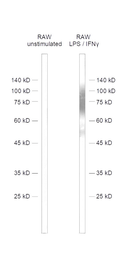
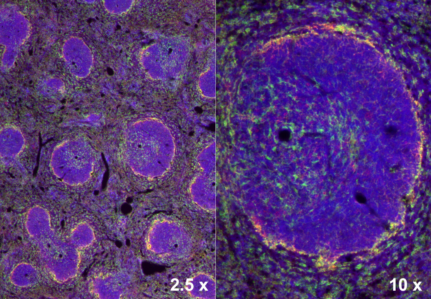
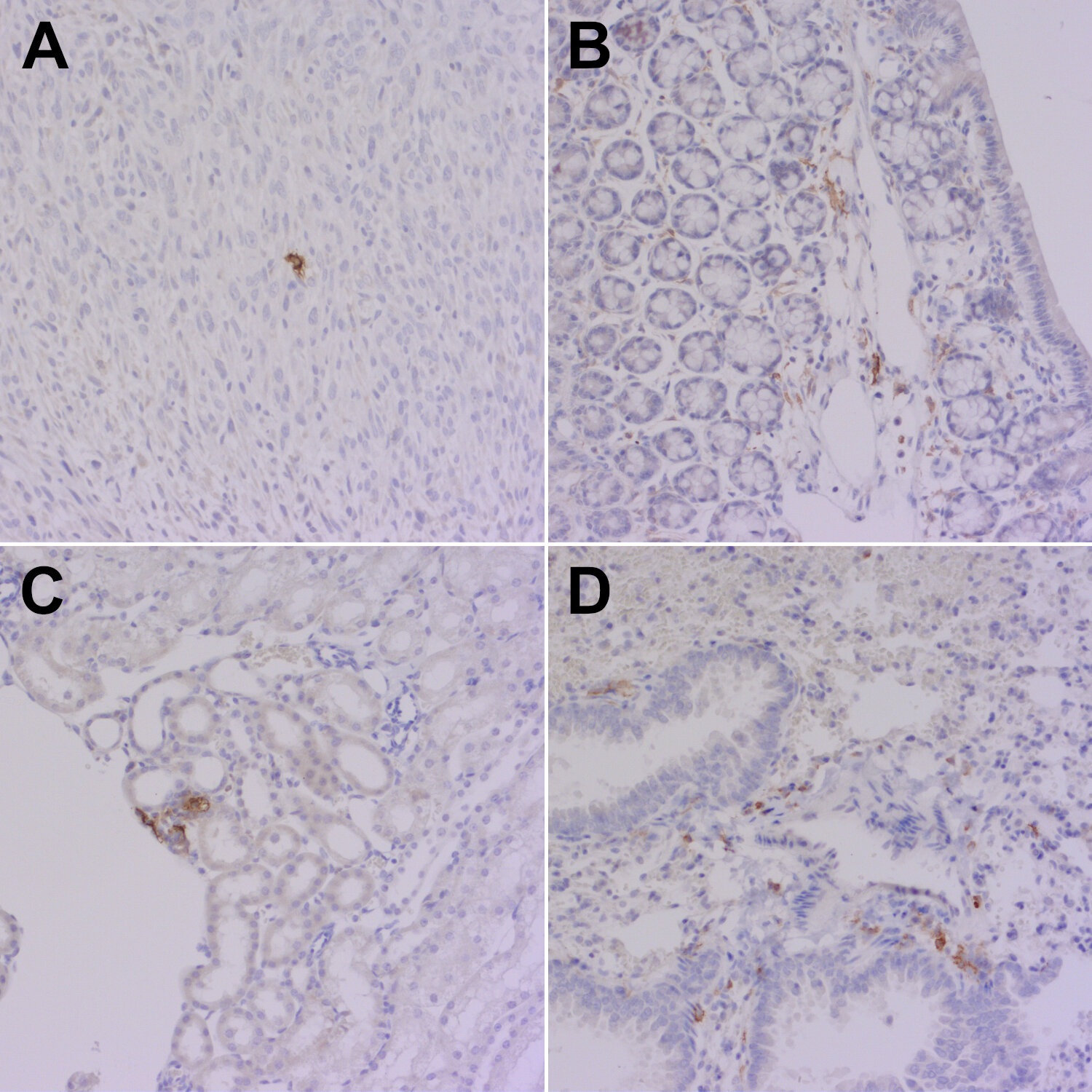
| Applications | |
| Immunogen | Synthetic peptide corresponding to AA 291 to 309 from mouse Cd86 (UniProt Id: P42082) |
| Reactivity |
Reacts with: mouse (P42082). No signal: human (P42081), rat. Other species not tested yet. |
| Remarks |
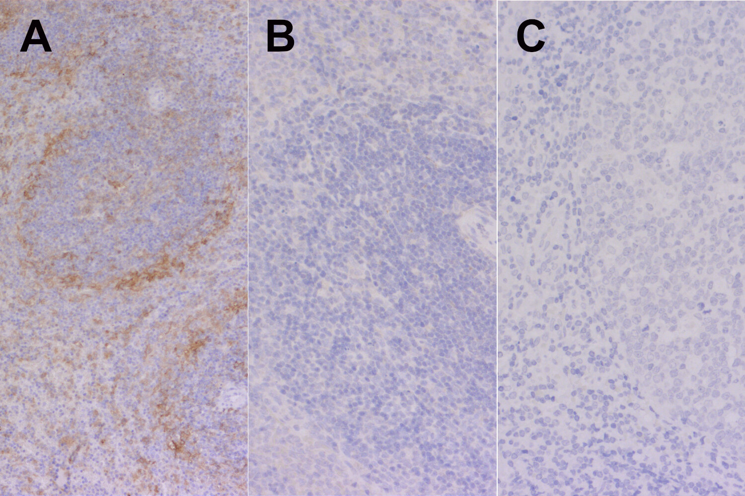
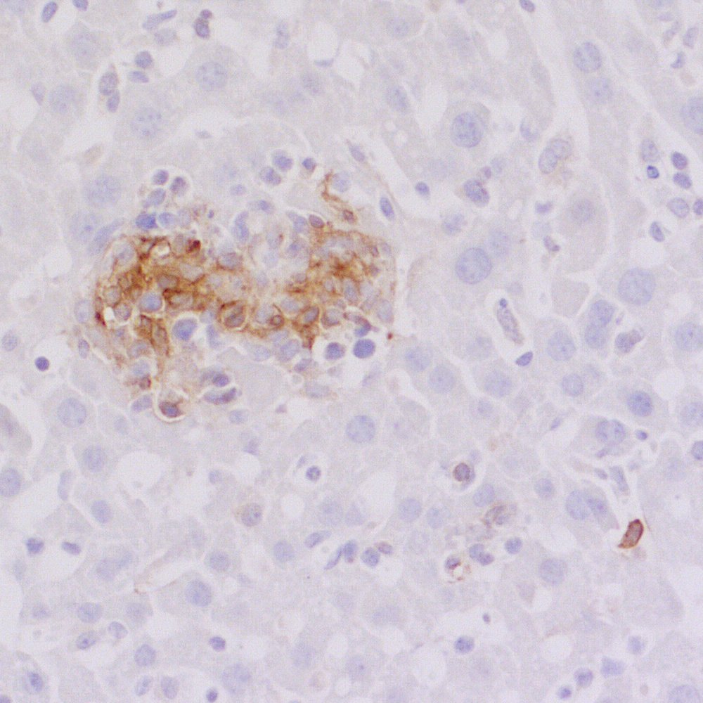
IHC: Antigen retrieval with citrate buffer pH 6 is required. |
| Data sheet | Datasheet hs-466_003 |
|
|
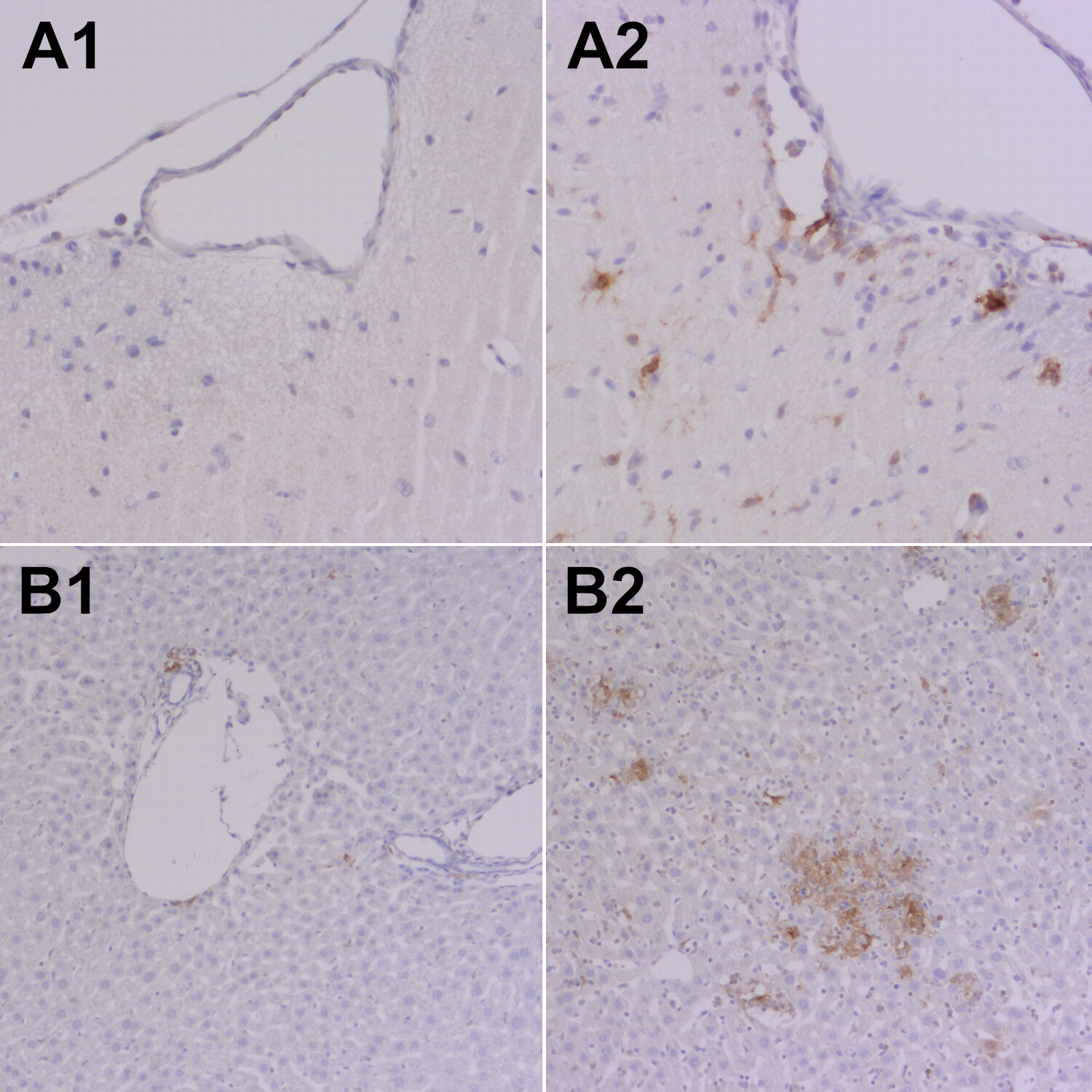
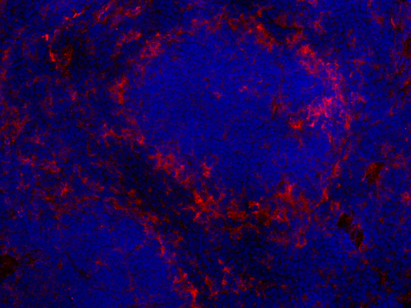
CD86 positive cells in a Toxoplasmose gondii infected mouse liver section
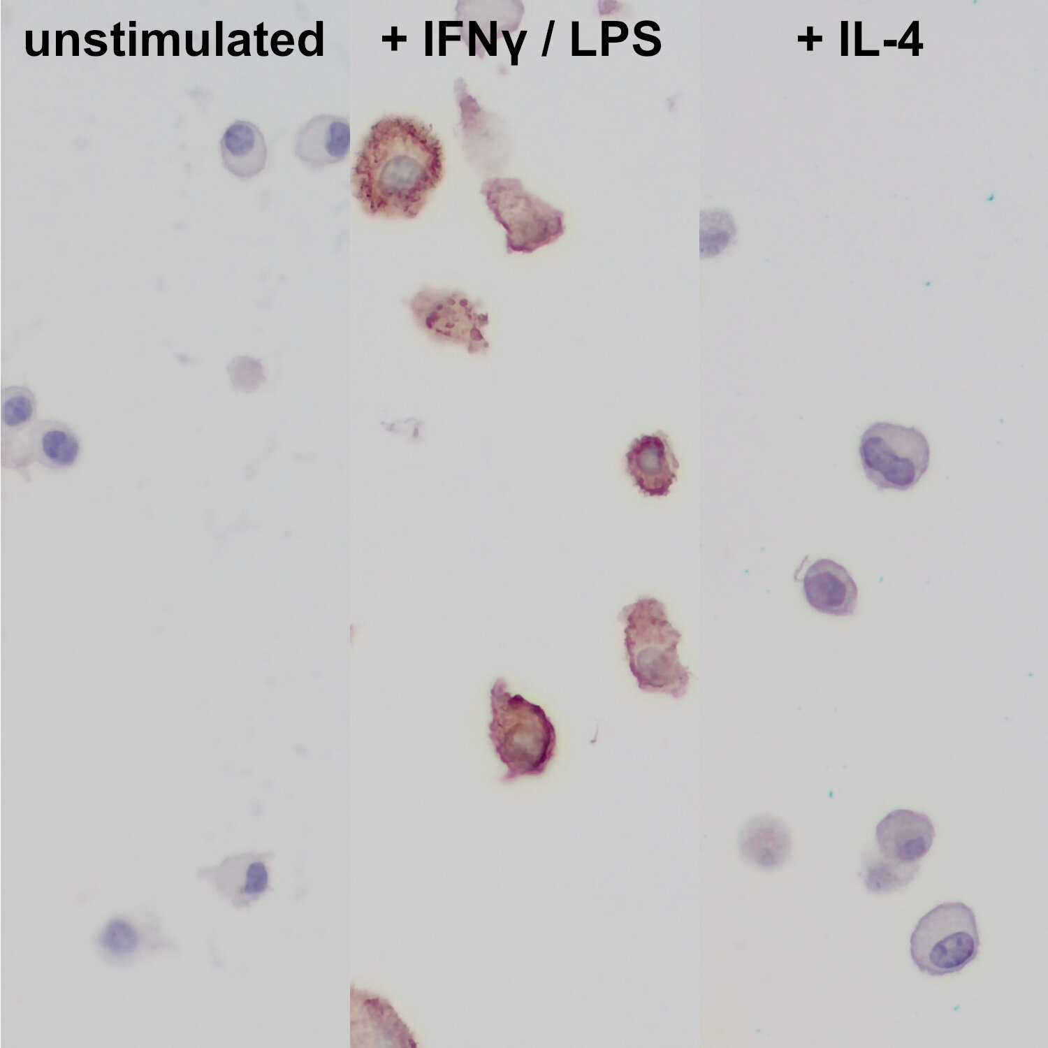
CD86 (Cluster of Differentiation 86, also known as B7.2) belongs to the B7 family of immune-regulatory cell-surface protein ligands (1). CD86 and the genetically closely linked CD80 protein (also known as B7.1) are expressed by antigen presenting cells and provide costimulatory signals necessary for T cell activation and tolerance via interaction with CD28 and cytotoxic T-lymphocyte antigen 4 (CTLA-4) expressed on T-cells. However, CD80 and CD86 have non-equivalent roles in immune modulation: CD86 is the dominant ligand for proliferation and survival of regulatory T cells (Tregs) (2) and shows in comparison with CD80 very high efficiency at increasing T cell killing capacity (3). CD86 is expressed only at low levels on resting B cells, dendritic cells and macrophages; activation results in enhanced CD86 expression (Collins et al., 2005). In the CNS, CD86 upregulation is a marker of activated pro-inflammatory M1 microglia (4). In oncology research, CD86 is a biomarker to phenotypically differentiate classically activated M1 macrophages from alternatively activated M2 macrophages in the tumor microenvironment (5).
Certificates
ISO 9001 2015 Quality Management System and Green Lab Platinum certification level for sustaining laboratory processes.


Newsletter
Sign up for our newsletter and get the latest updates and news.