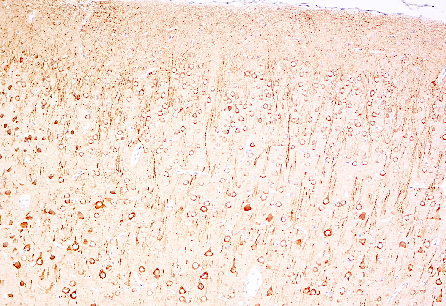
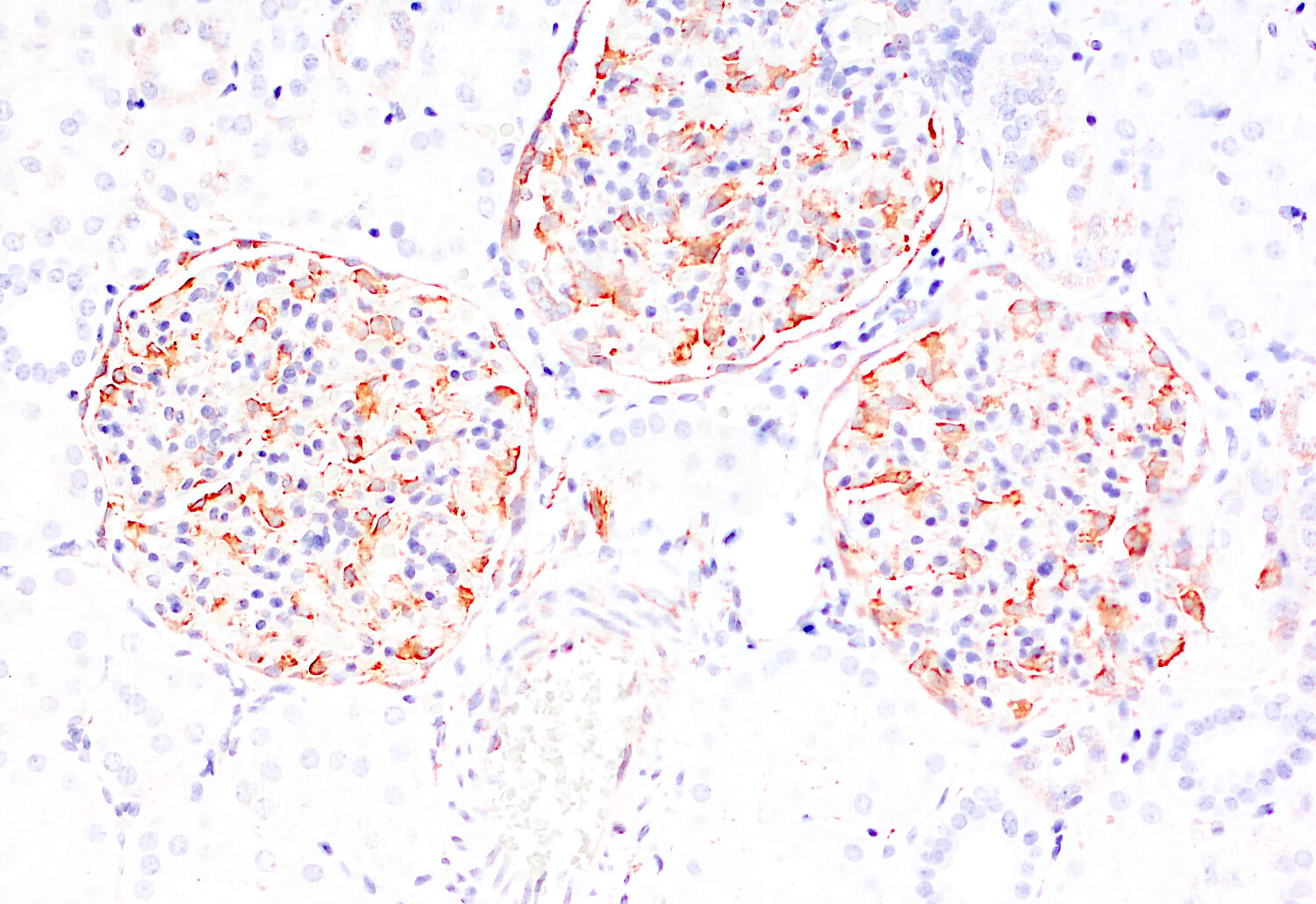
Cat. No.: 557 003
Amount: 50 µg
Price:
$380.00


|

|
|
| Cat. No. 557 003 |
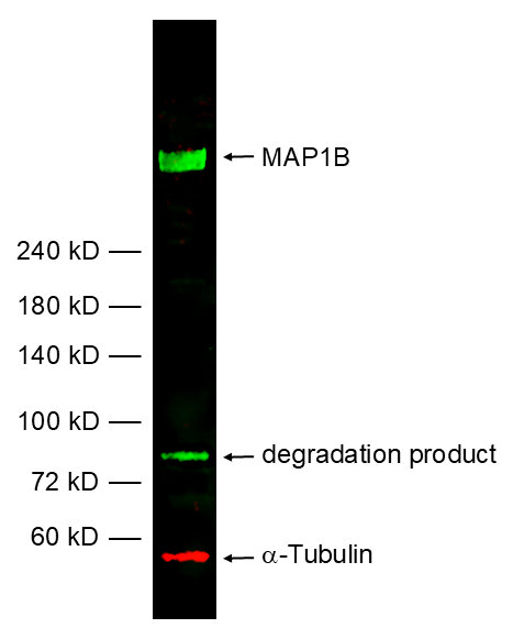
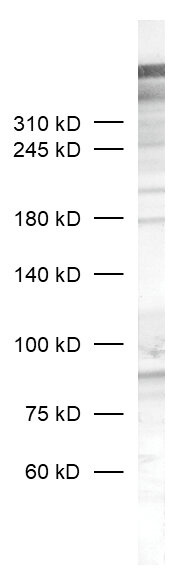
50 µg specific antibody, lyophilized. Affinity purified with the immunogen. Albumin and azide were added for stabilization. For reconstitution add 50 µl H2O to get a 1mg/ml solution in PBS. Then aliquot and store at -20°C to -80°C until use. Antibodies should be stored at +4°C when still lyophilized. Do not freeze! |
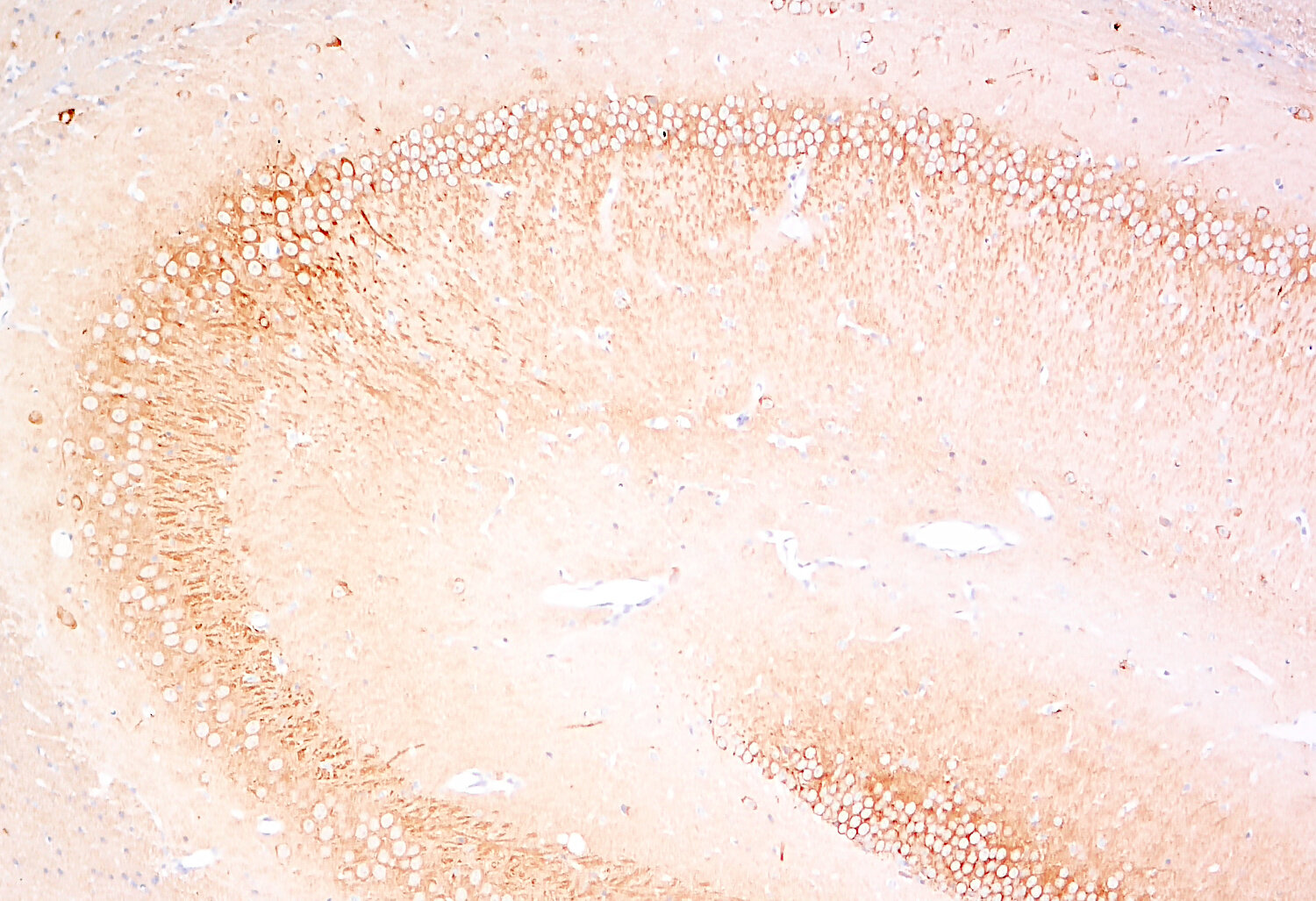
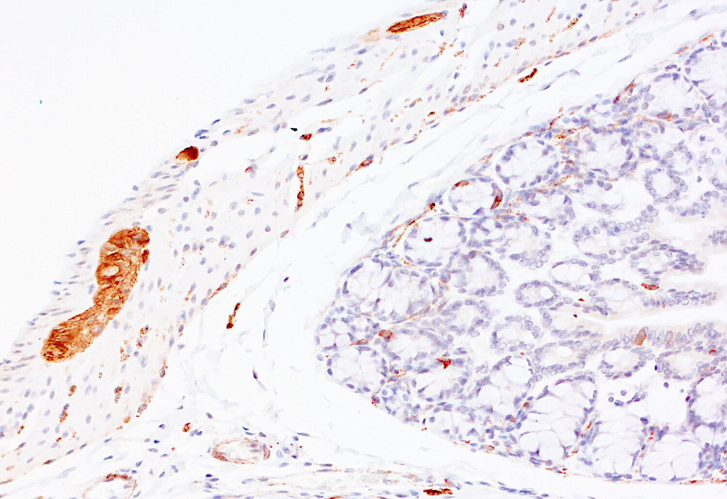
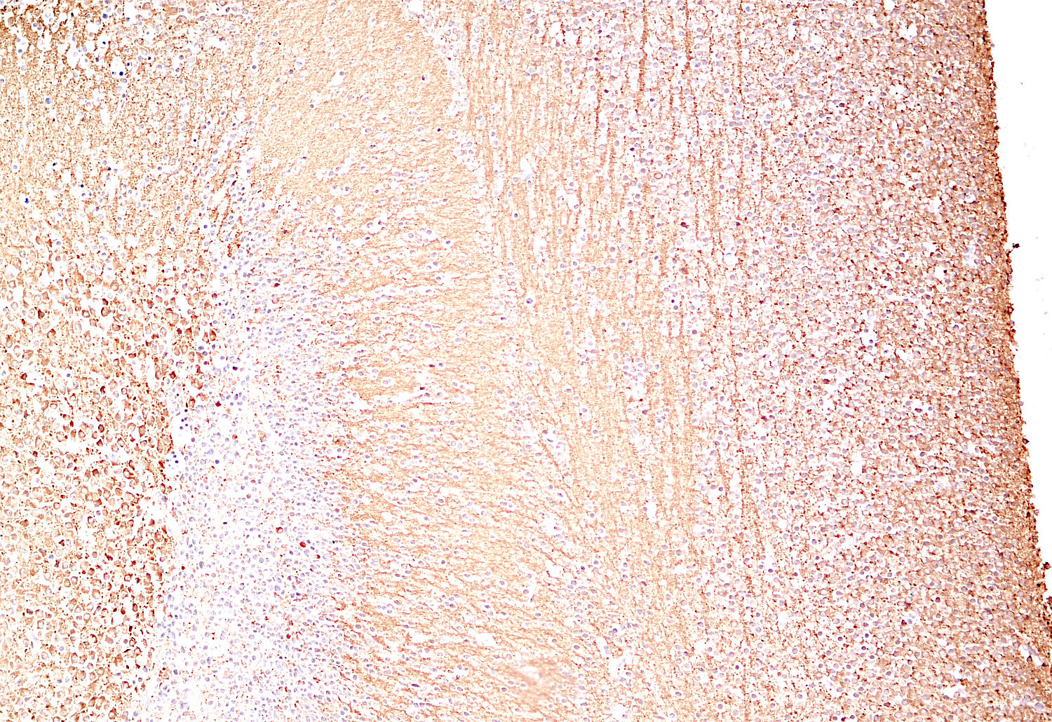
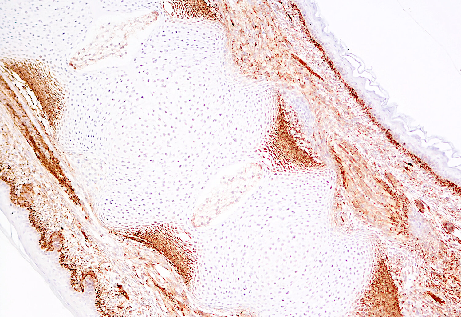
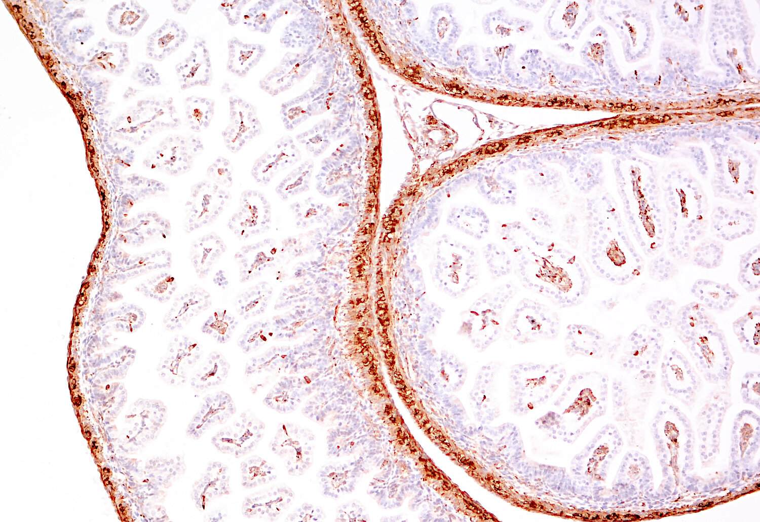
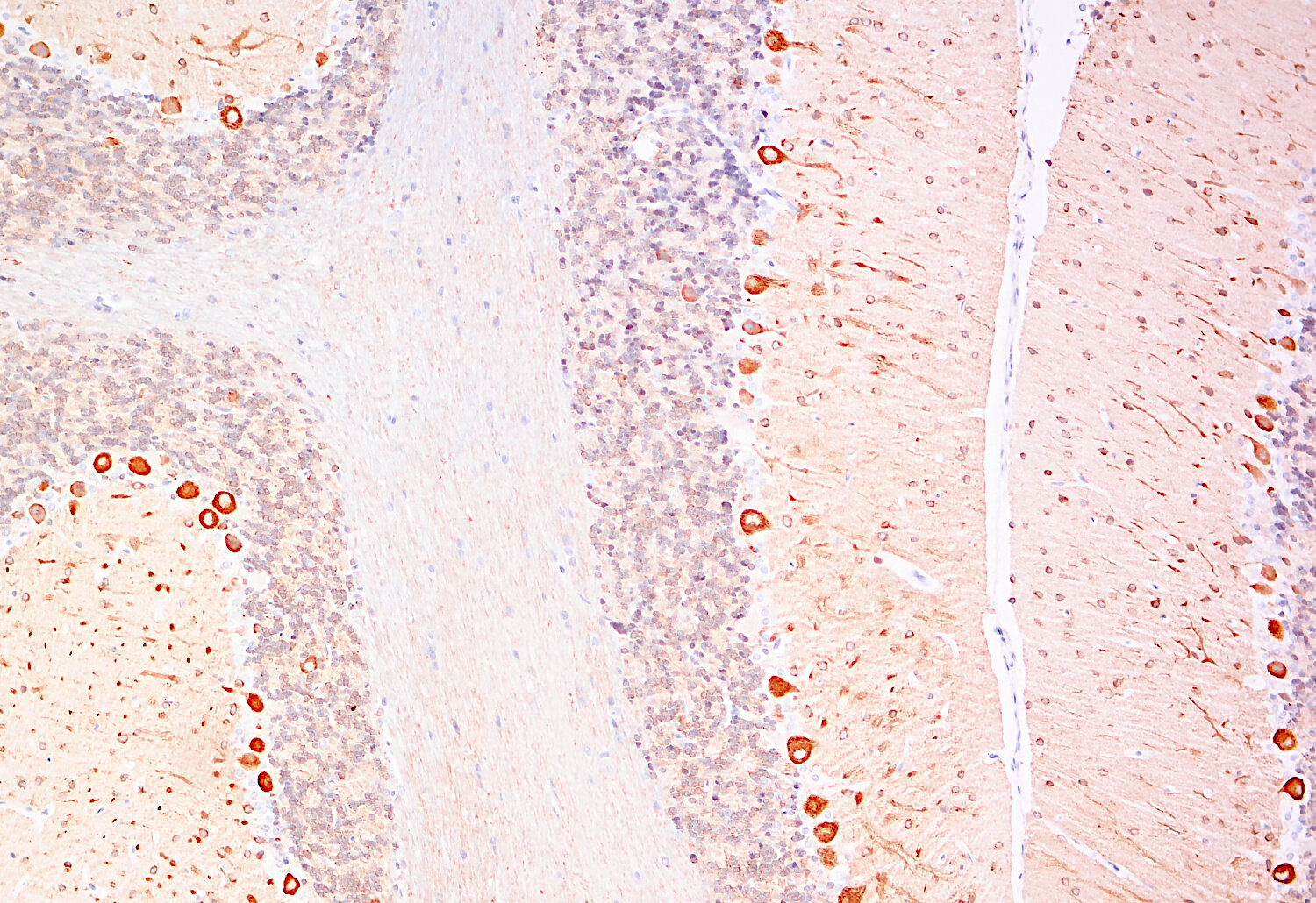
| Applications | |
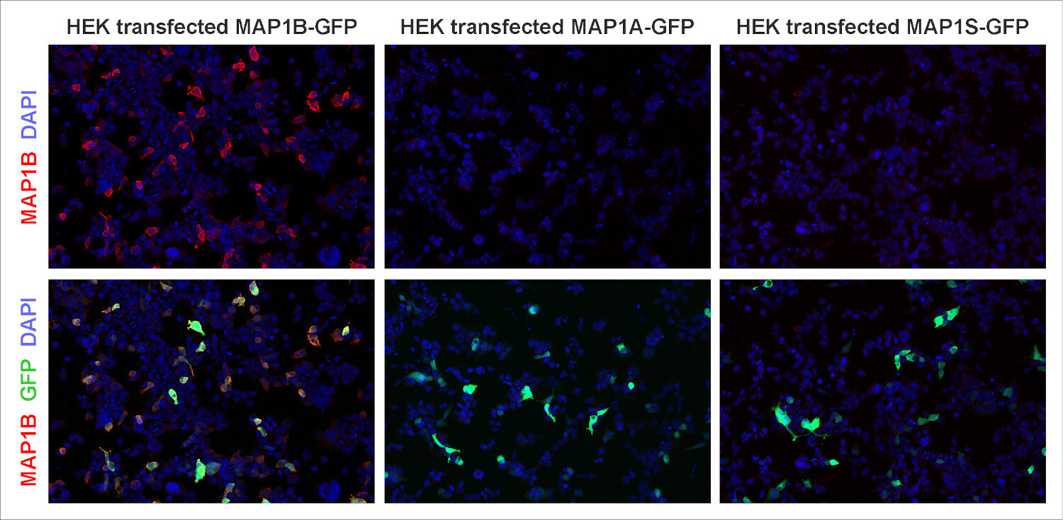
| Immunogen | Recombinant protein corresponding to the N-terminal part of mouse MAP1B (UniProt Id: P14873) |
| Reactivity |
Reacts with: mouse (P14873), rat (P15205), human (P46821). Other species not tested yet. |
| Data sheet | Data Sheet 557_003 |
|
|
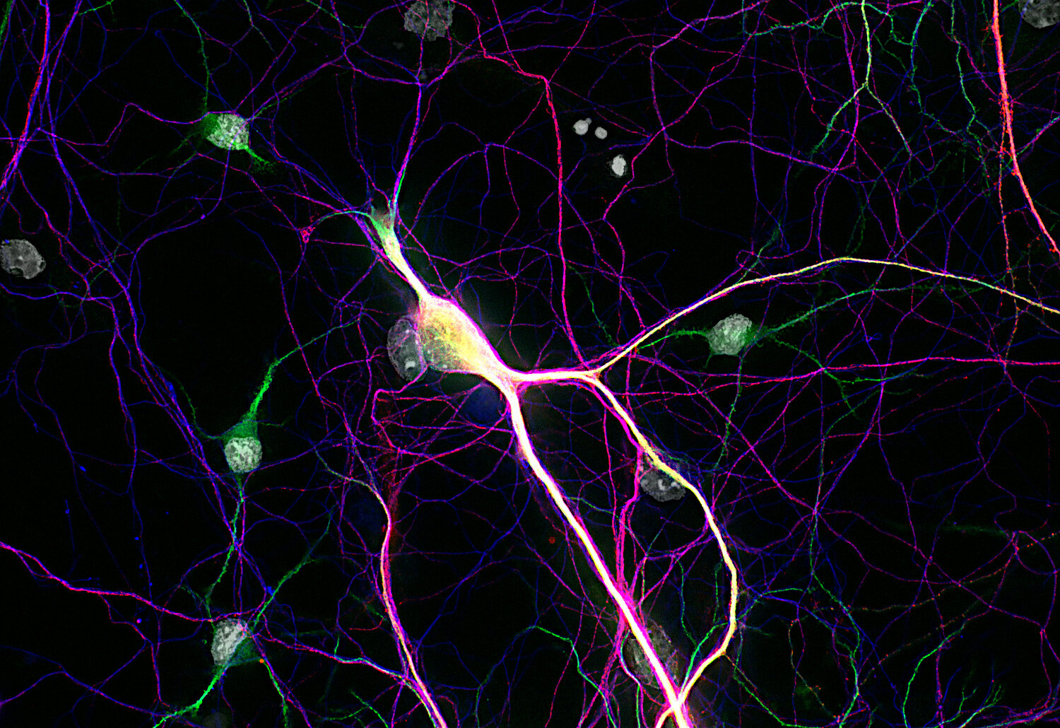
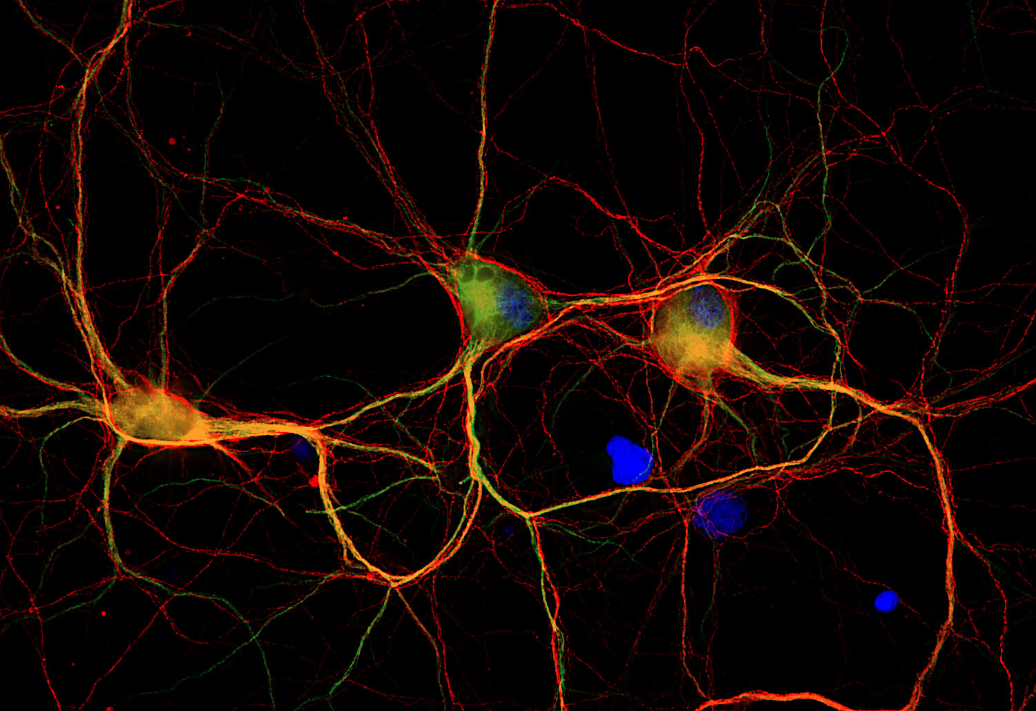
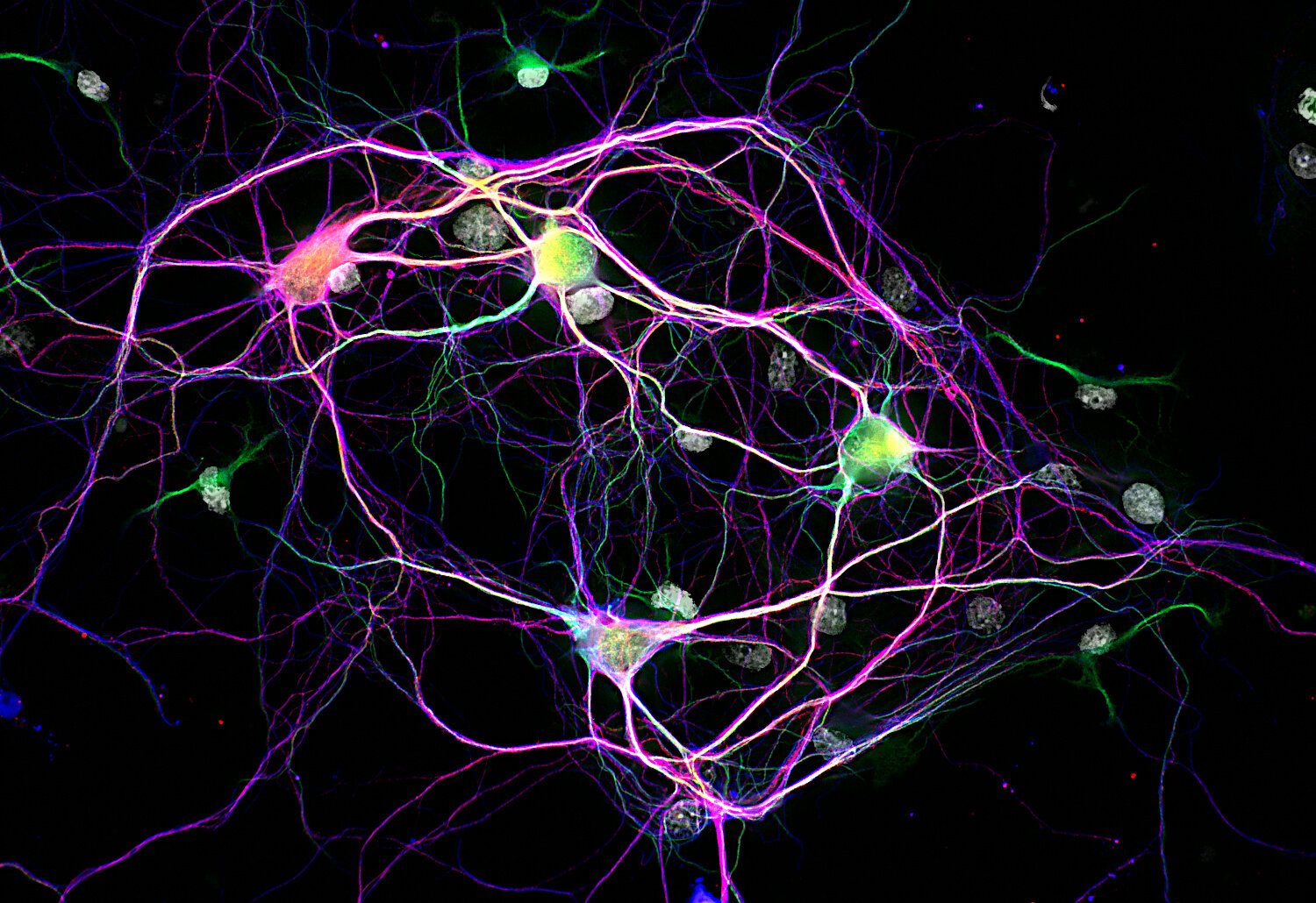
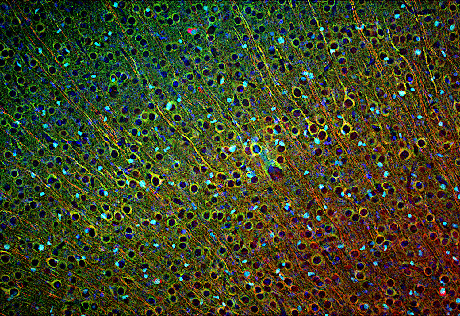
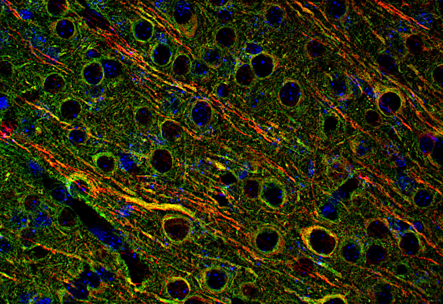
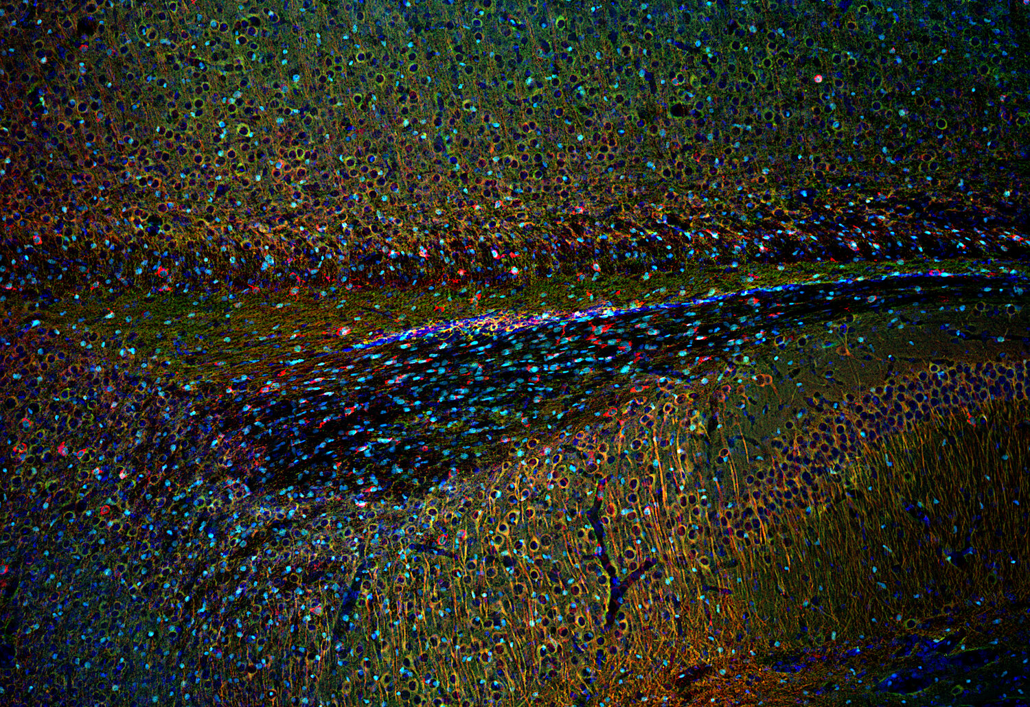
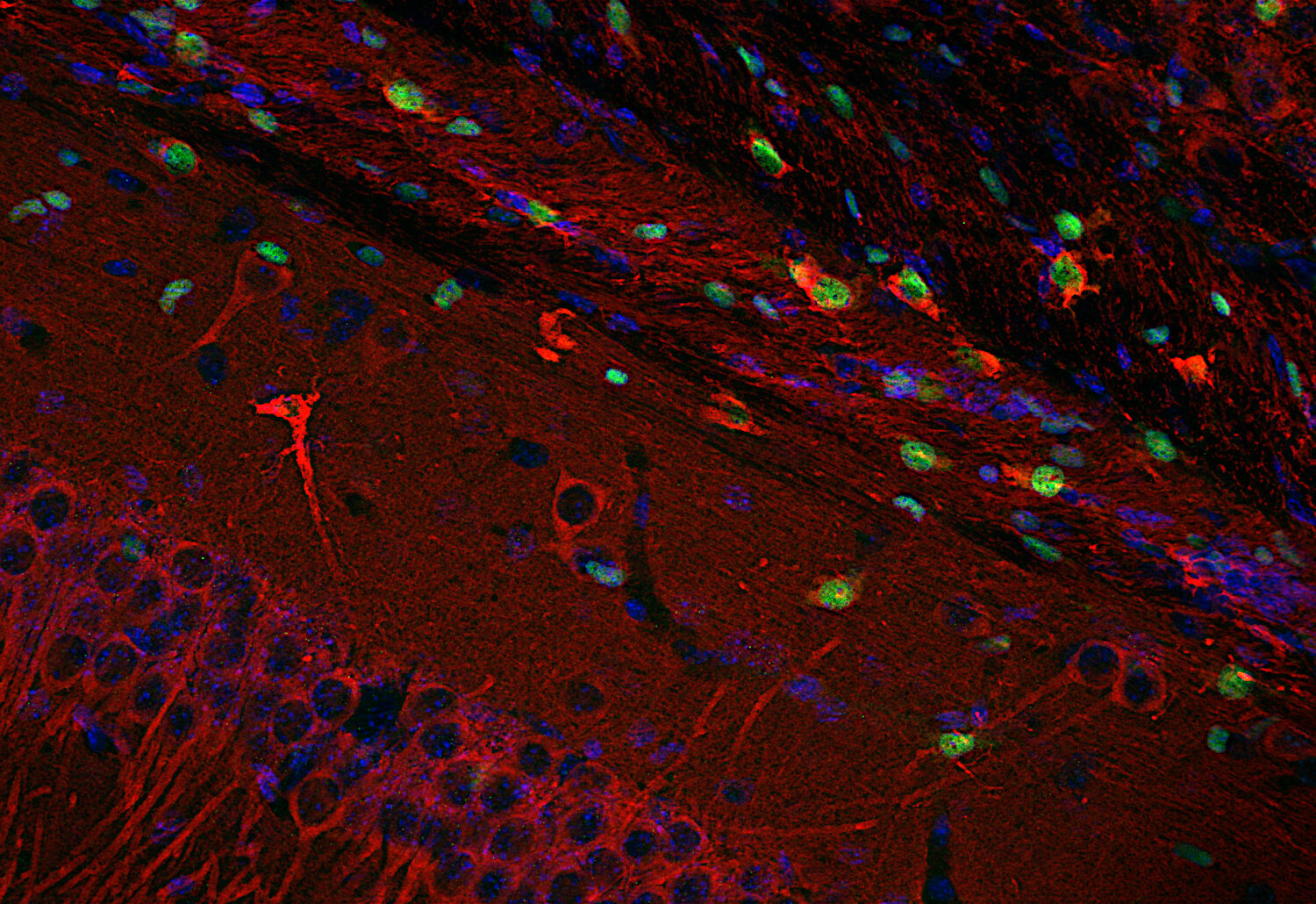
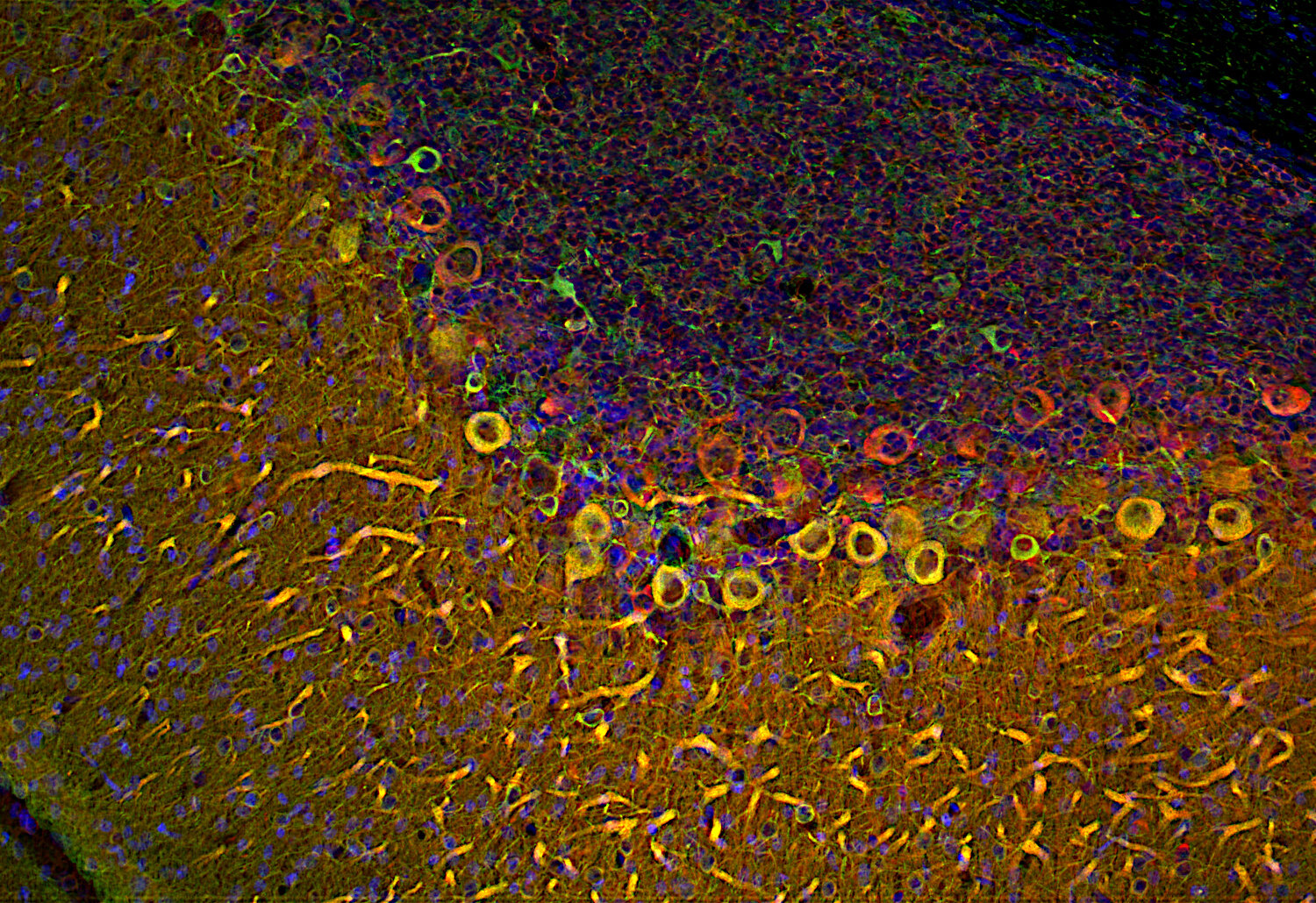
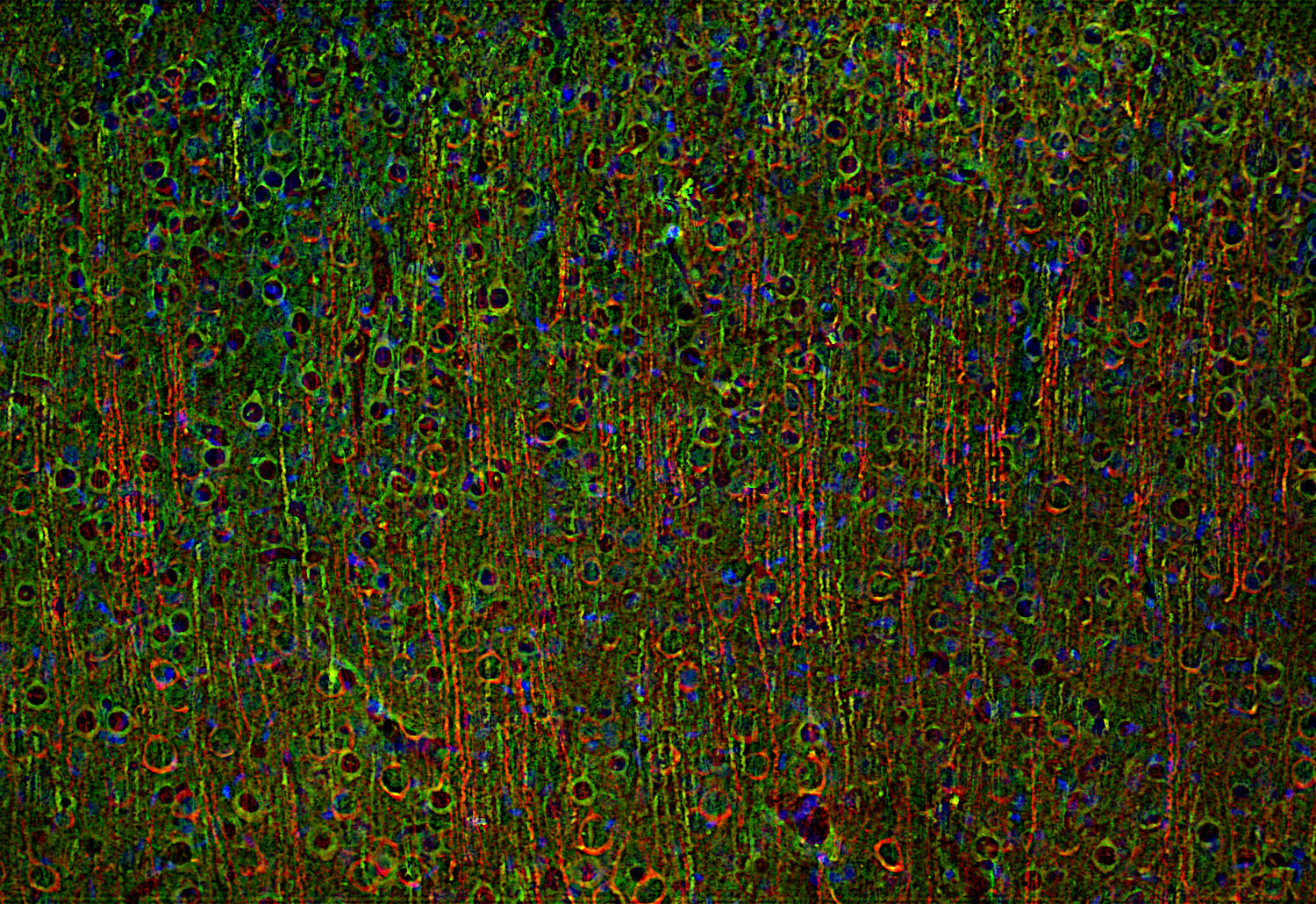
Rat hippocampus neurons stained for MAP1B (red), MAP2 (green) and beta3-Tubulin (blue)
Microtubule-associated protein 1B (MAP1B) is a neuronal microtubule-associated protein of the MAP1 family. It is synthesized as a precursor that is cleaved near the carboxy terminus into a heavy and a light chain which can interact with each other. MAP1B can bind microtubules and actin, linking the two cytoskeletal systems (1-3). The MAP1 protein family is conserved in vertebrates (2, 4).
MAP1B is expressed predominantly in the nervous system, especially during early development, where it is enriched in growing axons and growth cones, but it remains present in adult brain regions with high plasticity such as hippocampus, cortex, cerebellum, and olfactory bulb (2, 4, 5, 6).
Functionally, MAP1B regulates microtubule stability and actin dynamics, axon outgrowth and guidance, neuronal migration, dendritic spine development, synaptic maturation, and presynaptic organization (3, 5, 6, 7). MAP1B deficiency alters synaptic vesicle fusion, leads to abnormal presynaptic terminal structure and dendritic spines, and it impairs mitochondrial retrograde transport (5, 7, 8).
MAP1B also plays a role in the proliferation and migration of cancer cells, and human MAP1B mutations are correlated with the development of a variety of neurological diseases including Parkinson’s disease, schizophrenia, and periventricular nodular heterotopia (3).
Certificates
ISO 9001 2015 Quality Management System and Green Lab Platinum certification level for sustaining laboratory processes.

Newsletter
Sign up for our newsletter and get the latest updates and news.