Cat. No.: 551 003
Amount: 50 µg
Price:
$380.00
|
|

|
|
| Cat. No. 551 003 |
50 µg specific antibody, lyophilized. Affinity purified with the immunogen. Albumin and azide were added for stabilization. For reconstitution add 50 µl H2O to get a 1mg/ml solution in PBS. Then aliquot and store at -20°C to -80°C until use. Antibodies should be stored at +4°C when still lyophilized. Do not freeze! |
| Applications | |
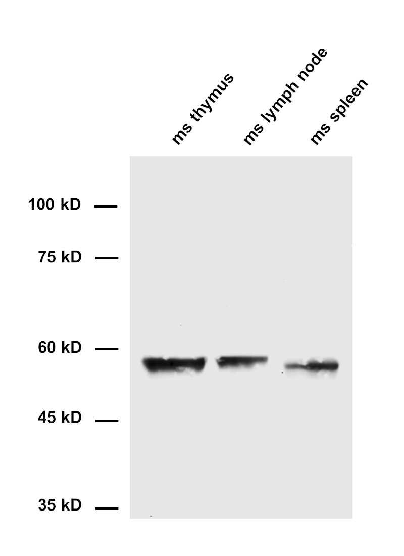
| Immunogen | Synthetic peptide corresponding to residues surrounding AA131 of mouse CD25 (UniProt Id: P01590) |
| Reactivity |
Reacts with: mouse (P01590). No signal: human (P01589), rat. Other species not tested yet. |
| Remarks |
IHC: Antigen retrieval with citrate buffer pH 6 is required. |
| Data sheet | Datasheet 551_003 |
|
|
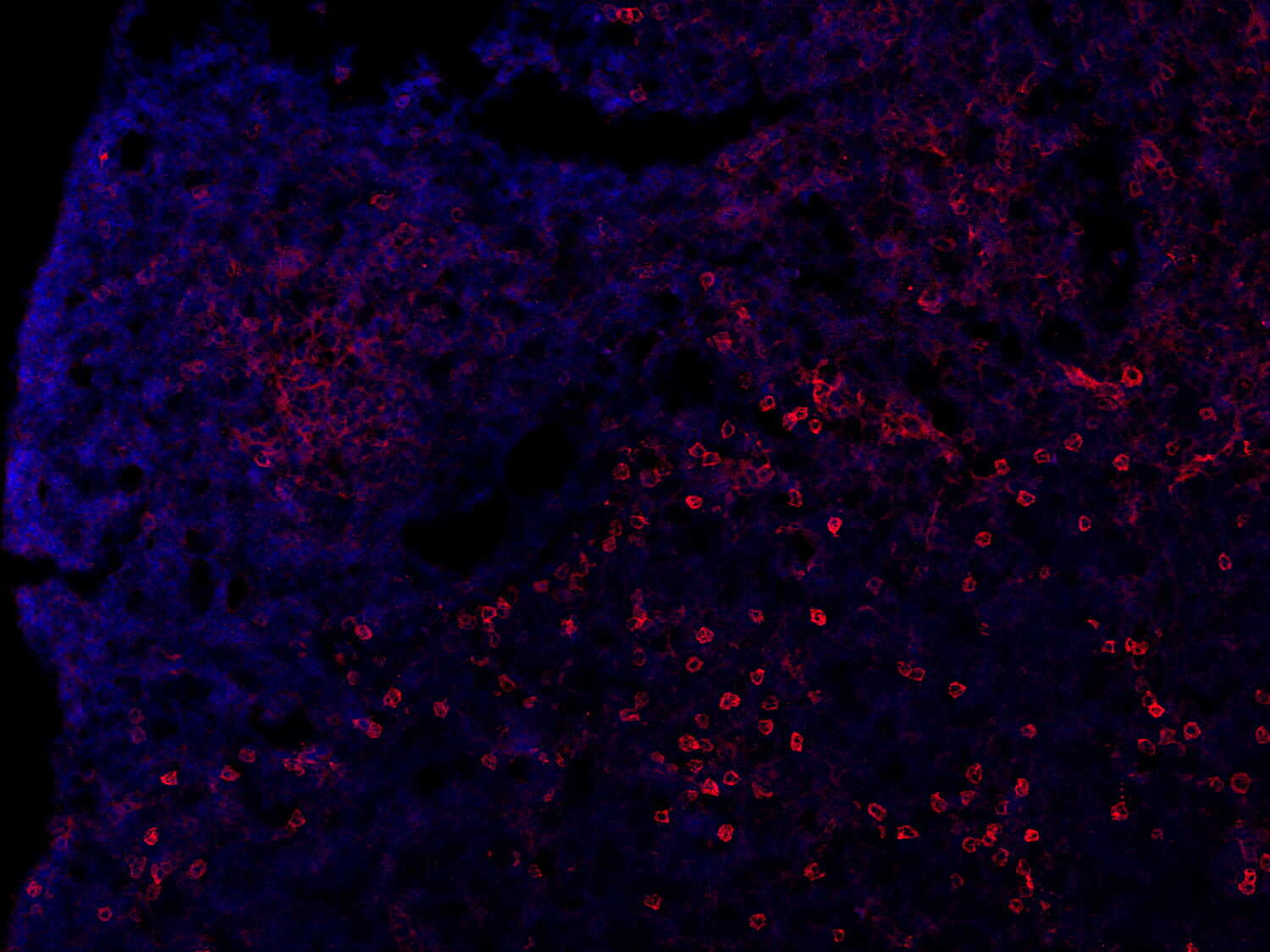
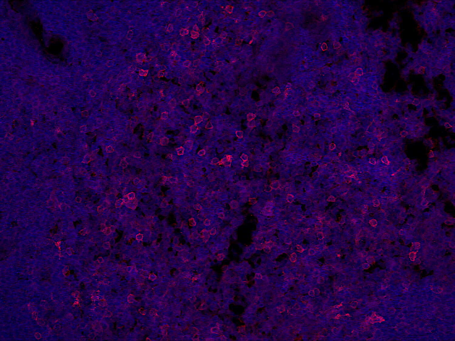
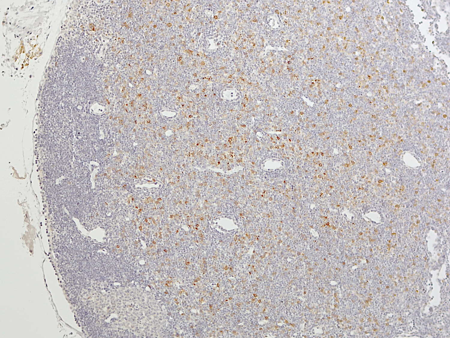
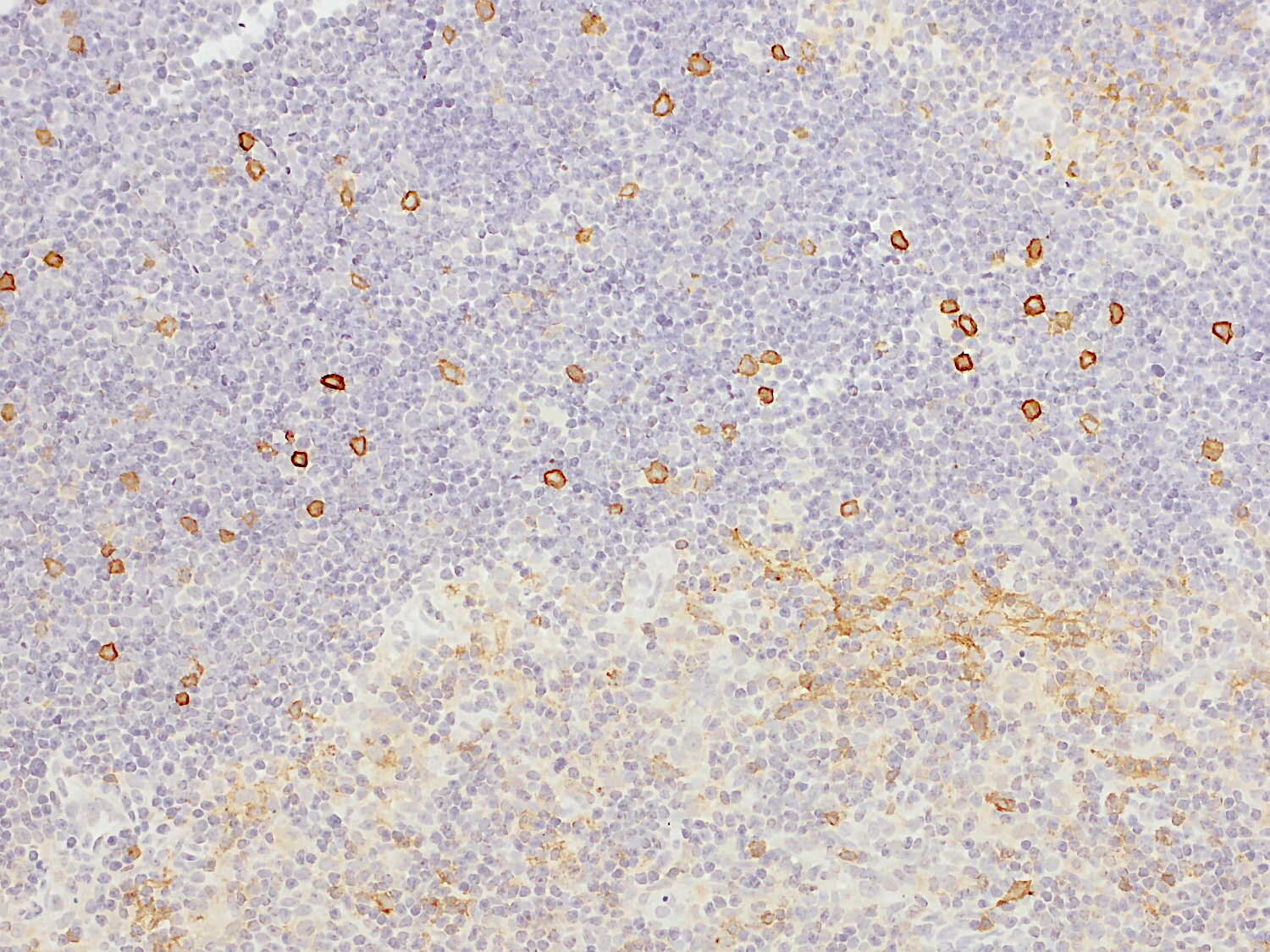
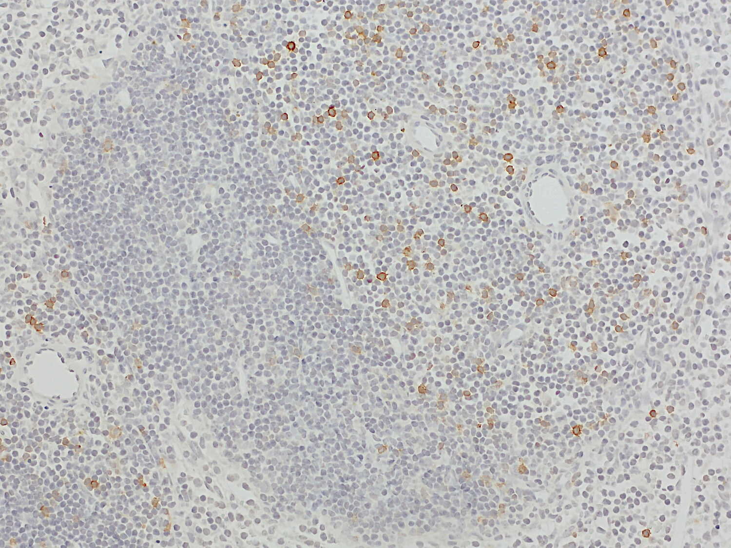
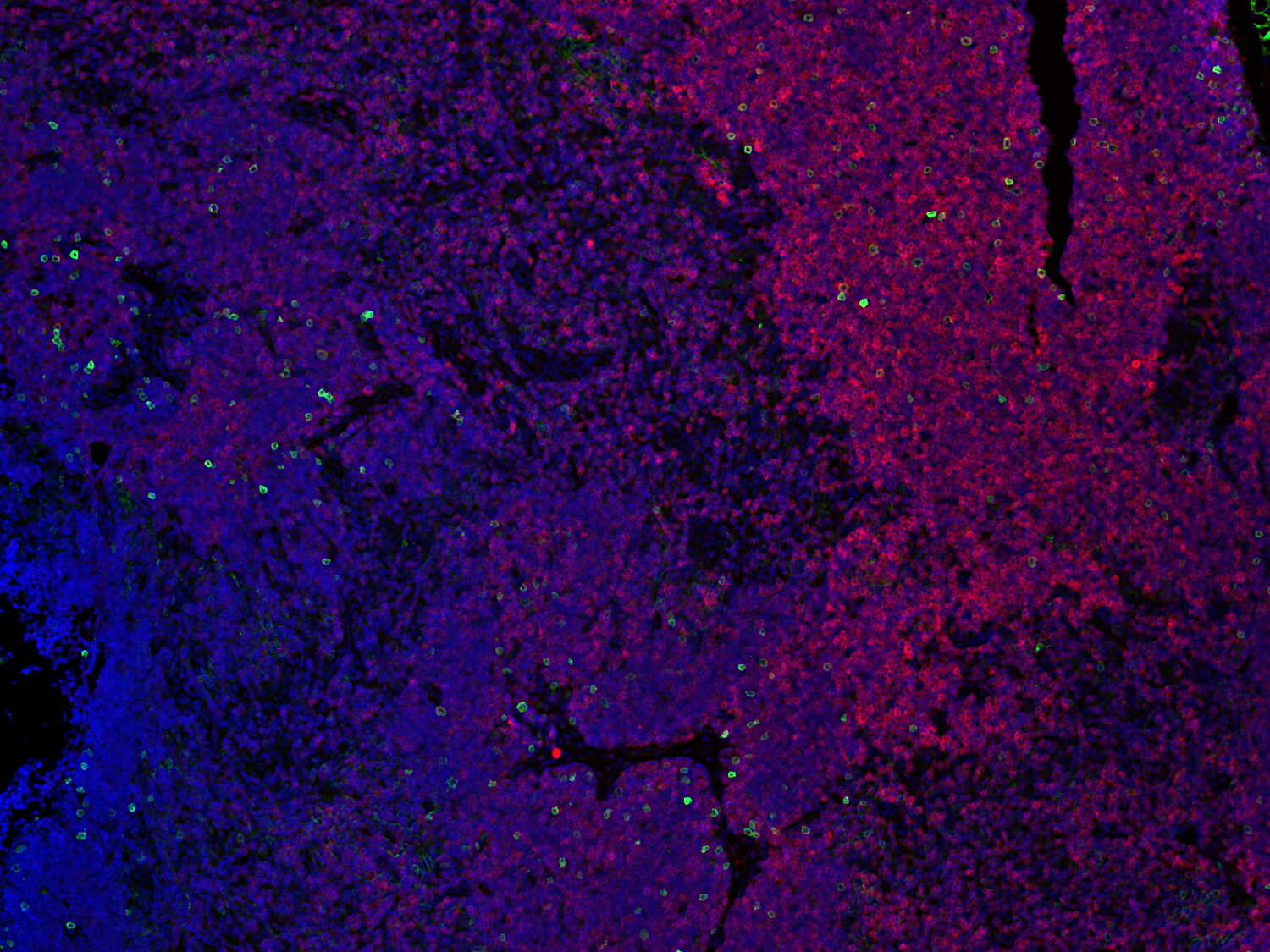
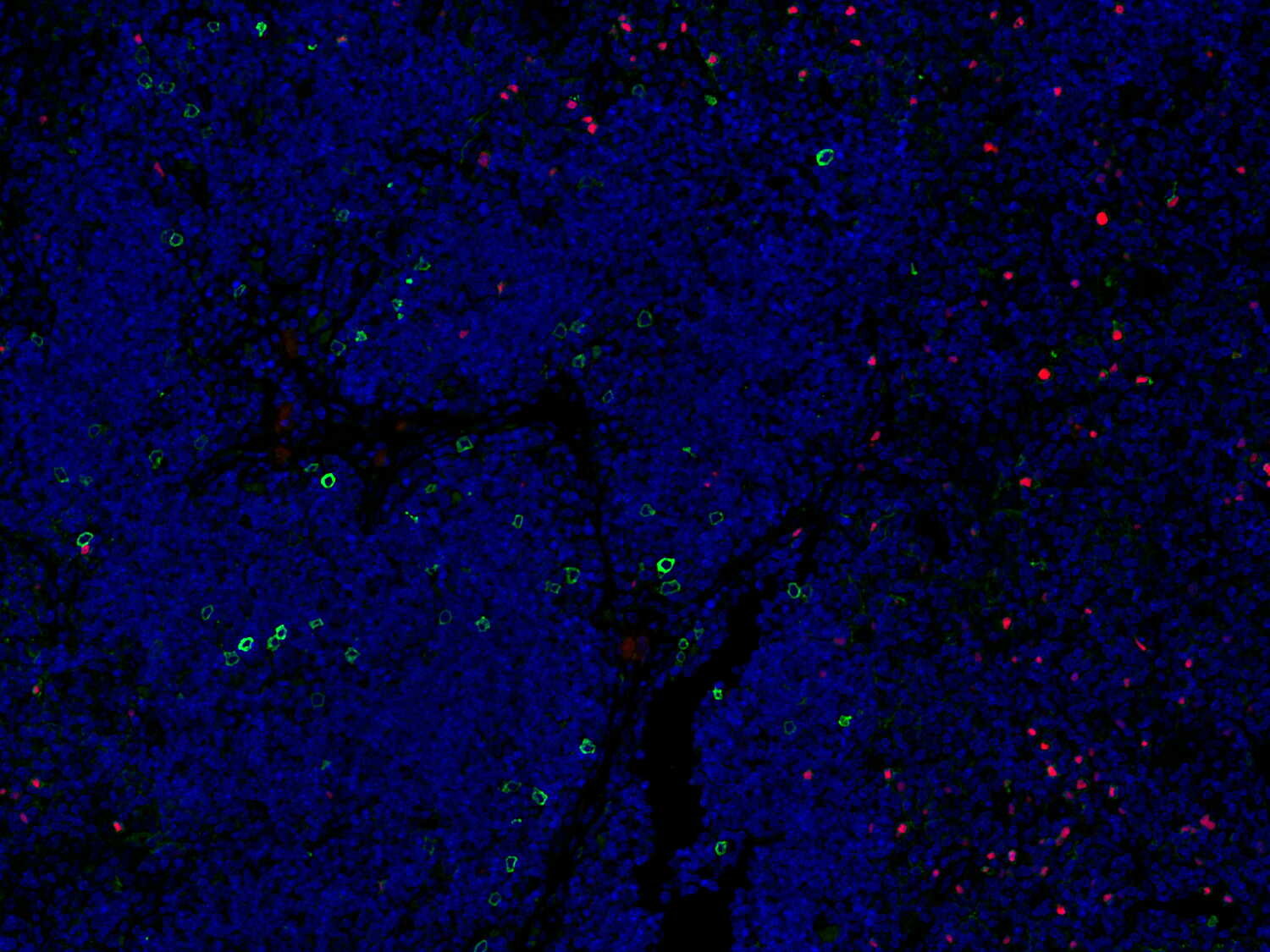
CD25 staining of a mouse thymus highlights early T cell progenitors and regulatory T cells
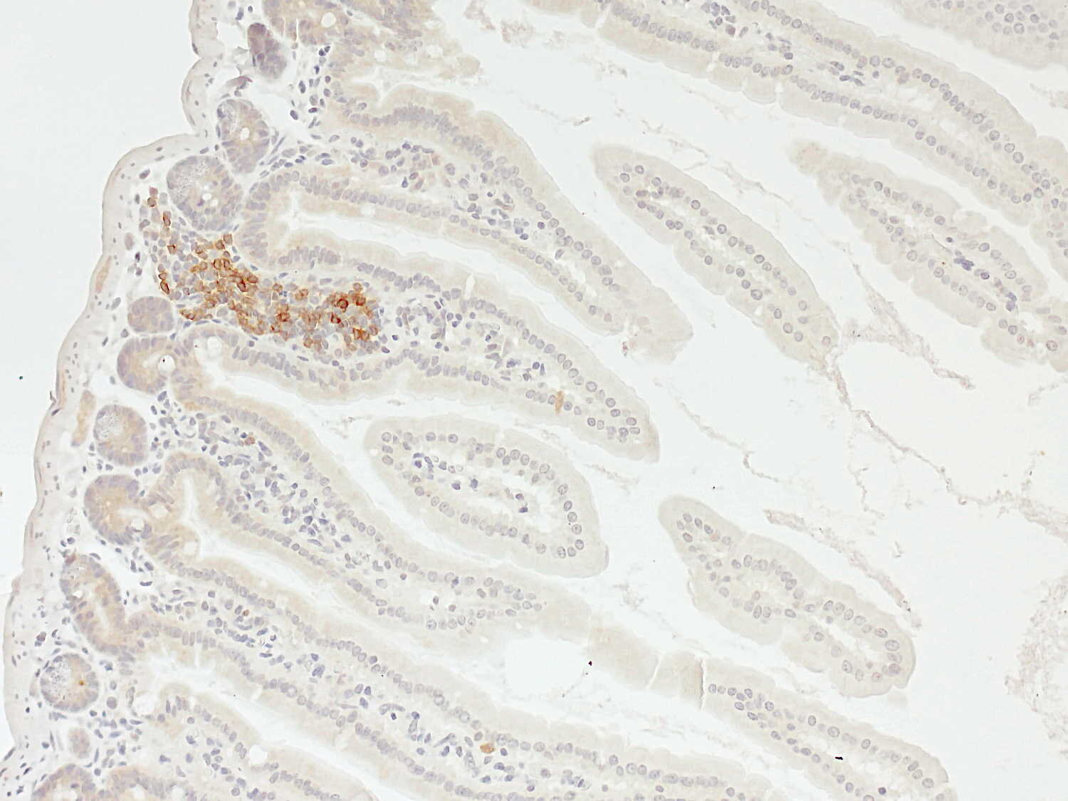
Interleukin-2 receptor alpha (IL-2Rα), also known as CD25, is a type I transmembrane glycoprotein that constitutes the α-chain of the trimeric interleukin-2 (IL-2) receptor complex. Structurally, CD25 comprises a large extracellular domain responsible for IL-2 binding, a single transmembrane region, and a short cytoplasmic tail that lacks intrinsic signaling motifs. Consequently, CD25 does not directly mediate intracellular signal transduction but primarily functions to increase the affinity of the IL-2 receptor for its ligand (1). During thymic T-cell development, CD25 expression marks intermediate CD4−CD8− double-negative thymocyte populations, particularly the DN2 and DN3 stages (2). CD25 is also constitutively expressed at variable levels on regulatory T cells (Tregs) (3,4). High CD25 expression on Foxp3⁺ Tregs is associated with potent immunosuppressive activity and has been linked to poor prognosis in colorectal cancer (5). In contrast, Foxp3⁺ Tregs residing in the small intestinal epithelium markedly downregulate CD25 expression and persist independently of IL-2 while retaining strong suppressive function (4).
Beyond its membrane-bound form, CD25 also exists as a soluble receptor generated through proteolytic shedding. Circulating levels of soluble CD25 are dysregulated in numerous pathological conditions, including cancer, inflammatory diseases, and autoimmune disorders, highlighting its relevance as a biomarker of immune activation (1).
Certificates
ISO 9001 2015 Quality Management System and Green Lab Platinum certification level for sustaining laboratory processes.


Newsletter
Sign up for our newsletter and get the latest updates and news.