Cat. No.: 523 002
Amount: 200 µl
Price:
$360.00


|

|
|
| Cat. No. 523 002 |
200 µl antiserum, lyophilized. For reconstitution add 200 µl H2O, then aliquot and store at -20°C until use. Antibodies should be stored at +4°C when still lyophilized. Do not freeze! |
| Applications | |
| Immunogen | Synthetic peptide corresponding to residues near the carboxy terminus of rat ATP13A5 (UniProt Id: F1MA70) |
| Reactivity |
Reacts with: human (Q4VNC0), rat (F1MA70). Other species not tested yet. |
| Remarks |
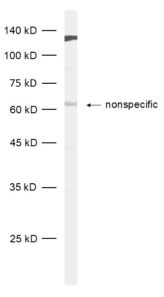
WB: recommended for Western blot. |
| Data sheet | Data Sheet 523_002 |
|
|
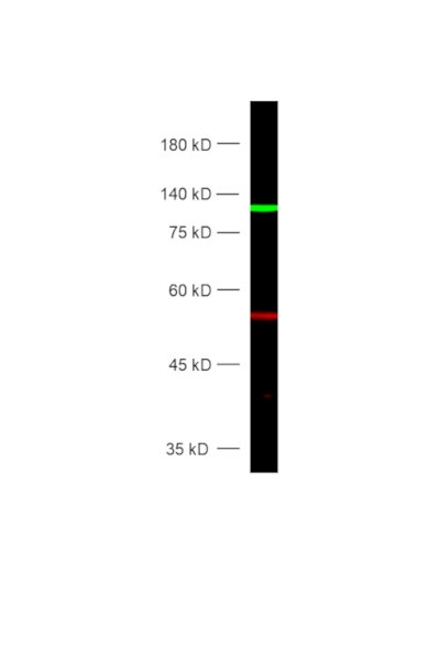
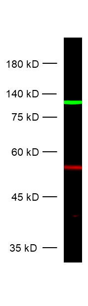
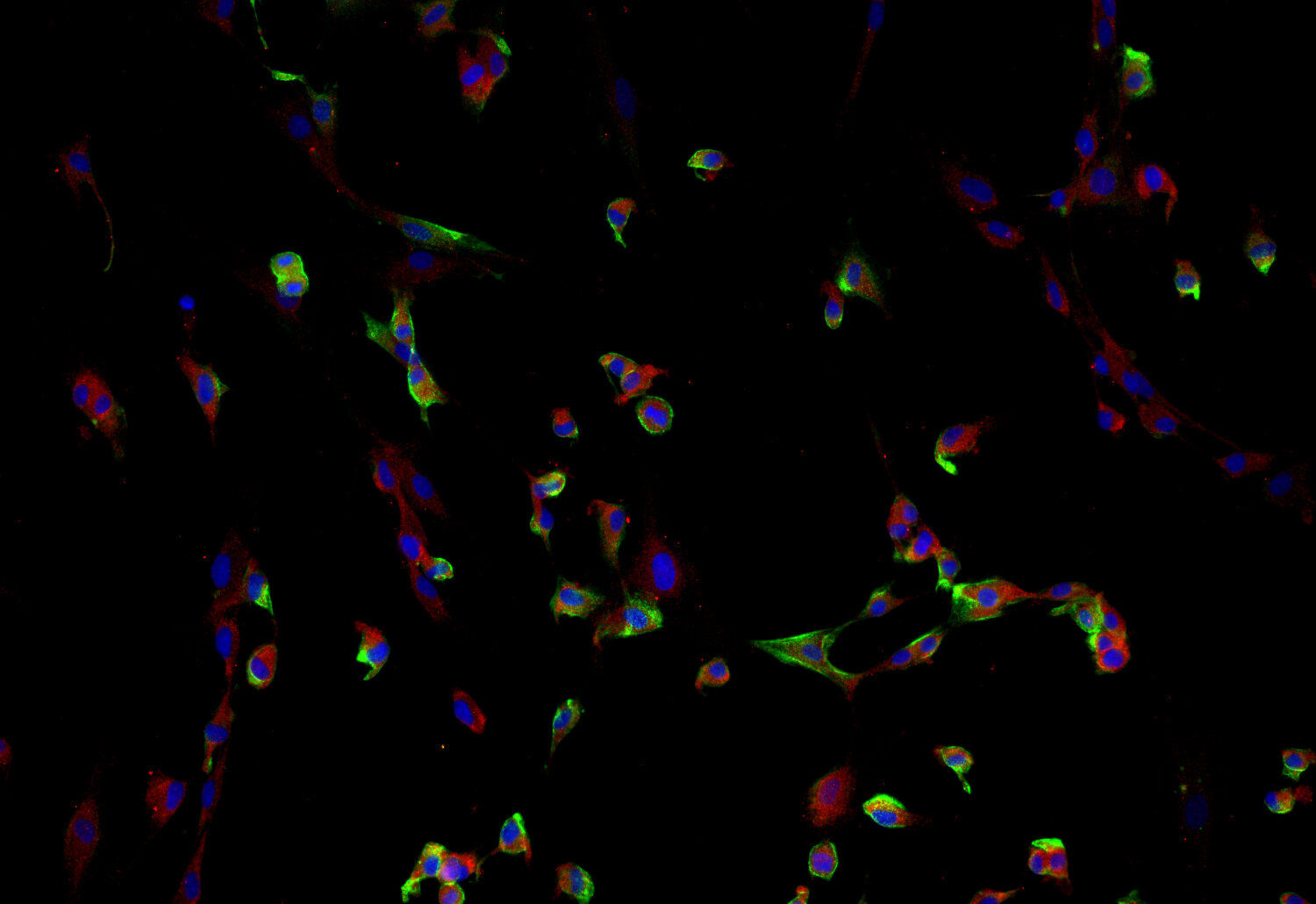
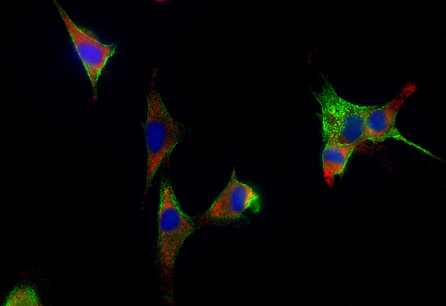
Detection of ATP13A5 (green) and alpha-Tubulin (red) in HeLa cell lysate
ATP13A5 is a marker that plays a key role in identifying central nervous system (CNS) pericytes, which are essential for vascular development and the maintenance of the blood-brain barrier (BBB). CNS pericytes are distinct from those in peripheral organs, and ATP13A5 has emerged as a specific genetic marker for these cells, validated through advanced transcriptomic and genetic models.
In mice, ATP13A5 expression is observed from embryonic day 15, aligning with the establishment of the BBB, and persists into adulthood, underscoring its role in CNS vasculature development. A knock-in model with ATP13A5-driven tdTomato reporter and Cre recombinase demonstrates that ATP13A5 expression is confined to CNS pericytes, including those in the brain, spinal cord, and retina, while showing minimal expression in peripheral tissues.
This marker enables precise genetic manipulation and detailed study of pericyte biology, including their development, heterogeneity, and function within the BBB. The specificity of ATP13A5 facilitates research into its role in neurological disorders, particularly those involving BBB dysfunction, such as Alzheimer's disease. The ATP13A5 model also supports the development of targeted therapies and genetic tools for studying CNS vascular health and disease (1).
Certificates
ISO 9001 2015 Quality Management System and Green Lab Platinum certification level for sustaining laboratory processes.


Newsletter
Sign up for our newsletter and get the latest updates and news.