Cat. No.: 314 008
Amount: 50 µg
Price:
$420.00
|
|

|
|
| Cat. No. 314 008 |
50 µg purified recombinant IgG, lyophilized. Albumin and azide were added for stabilization. For reconstitution add 50 µl H2O to get a 1mg/ml solution in PBS. Then aliquot and store at -20°C to -80°C until use. Antibodies should be stored at +4°C when still lyophilized. Do not freeze! |
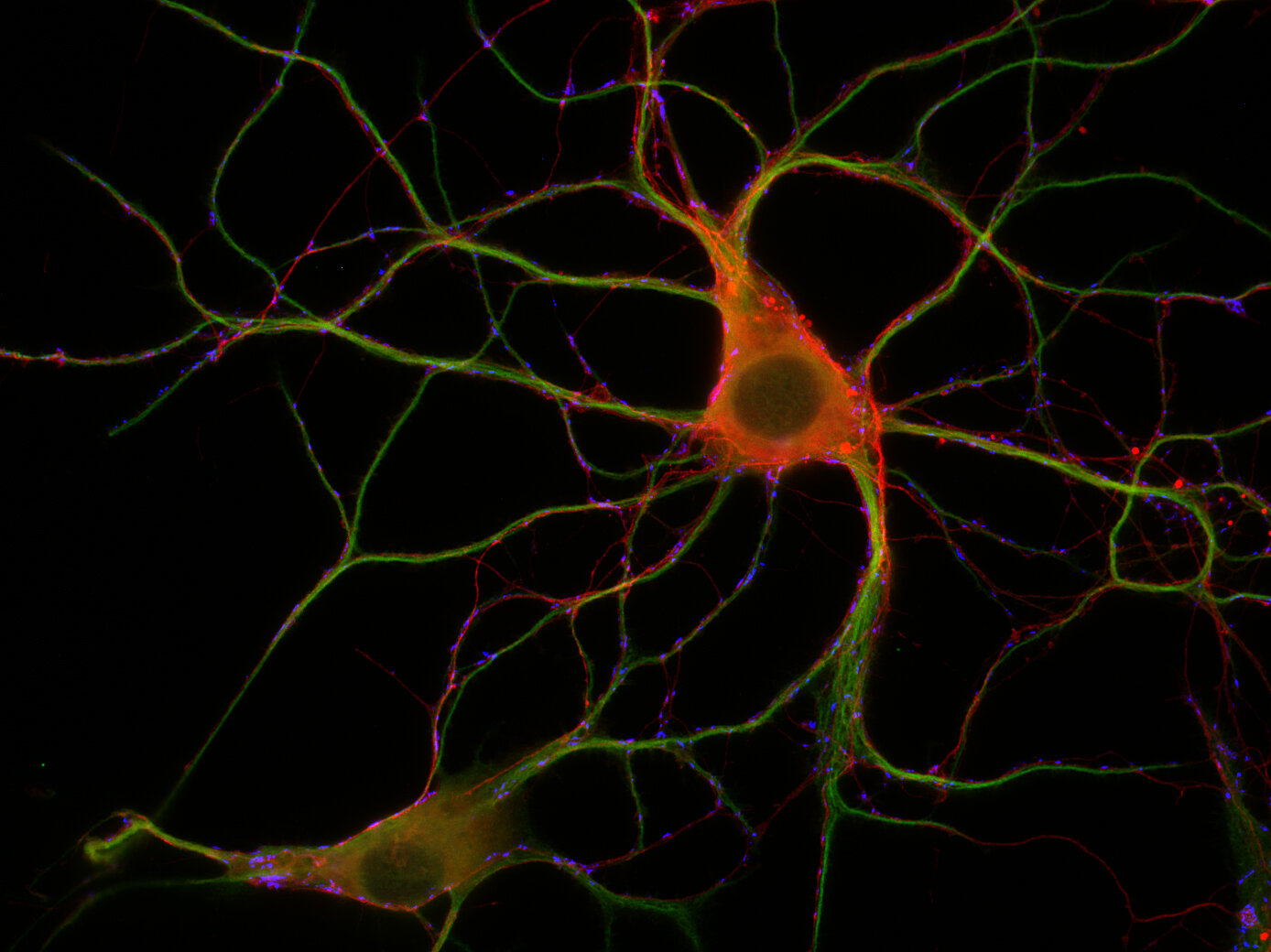
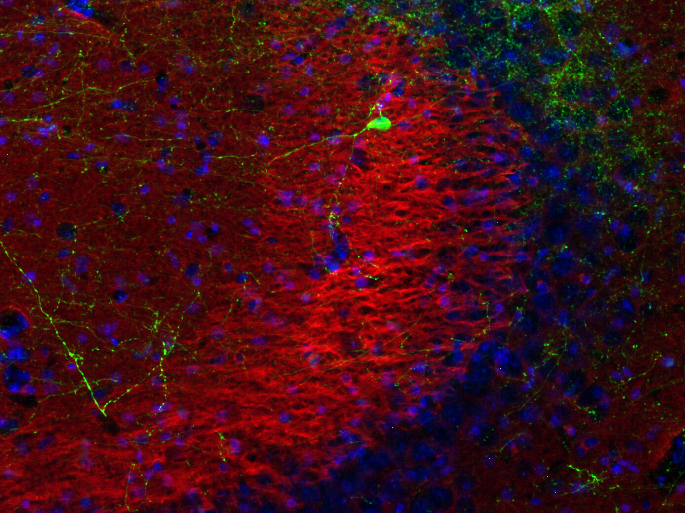
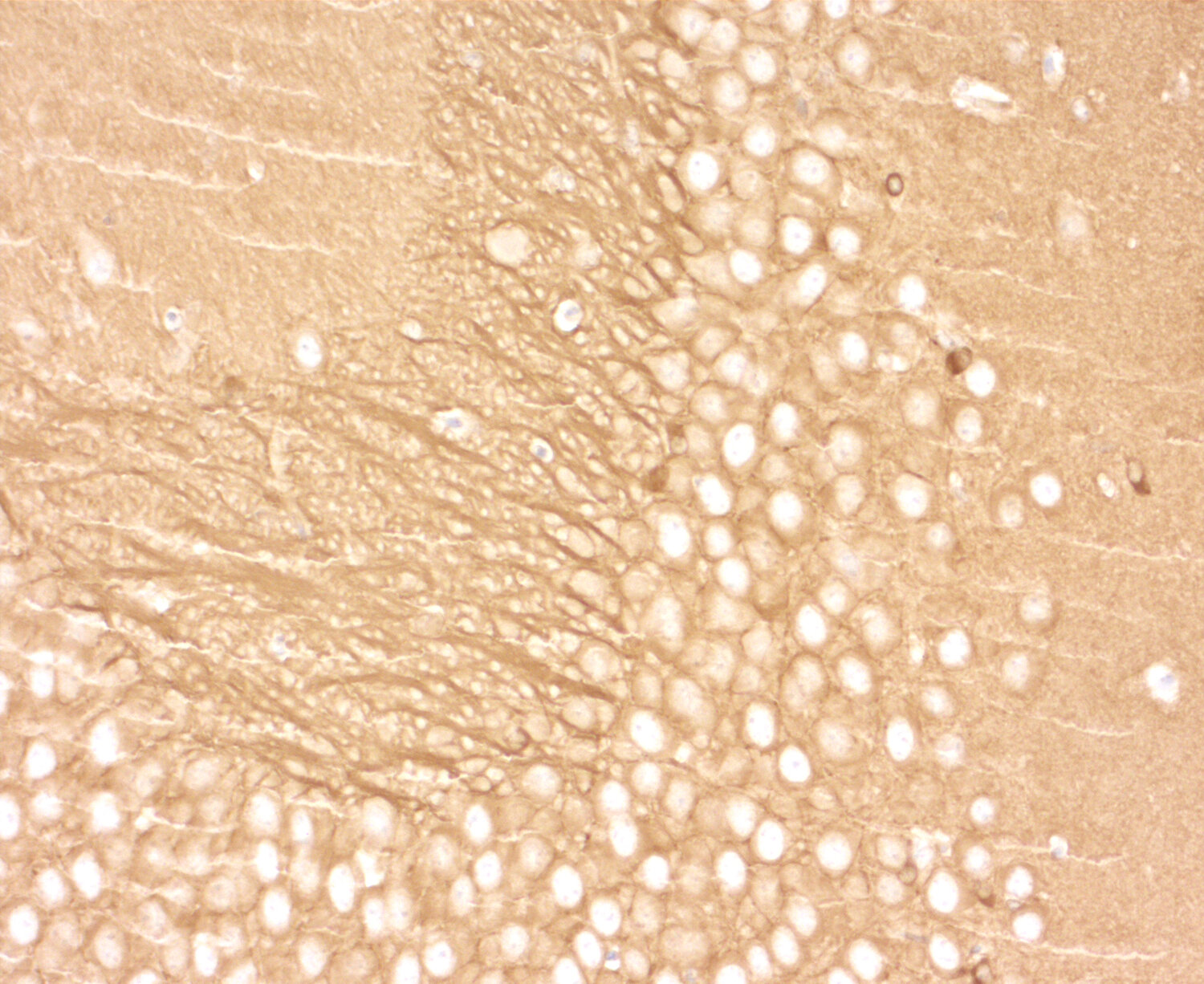
| Applications | |
| Clone | Rb248E5 |
| Subtype | IgG1 (κ light chain) |
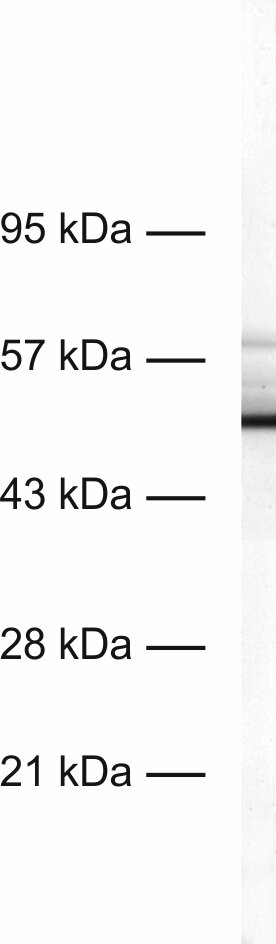
| Immunogen | Recombinant protein corresponding to the N-terminal half of mouse Tau-D (UniProt Id: P10637-5) |
| Reactivity |
Reacts with: rat (P19332), mouse (P10637). Weaker signal: human (P10636). No signal: zebrafish. Other species not tested yet. |
| Specificity | This antibody binds phosphorylated and non-phosphorylated tau proteins. The sequence used for immunization is present in all splice variants except human TauA (UniProt Id: P10636-3). |
| Matching control protein/peptide | 314-0P |
| Remarks |
This antibody is a chimeric antibody based on the monoclonal mouse antibody clone 248E5. The constant regions of the heavy and light chains have been replaced by rabbit specific sequences. Therefore, the antibody can be used with standard anti-rabbit secondary reagents. The antibody has been expressed in mammalian cells. |
| Data sheet | Datasheet 314_008 |
|
|
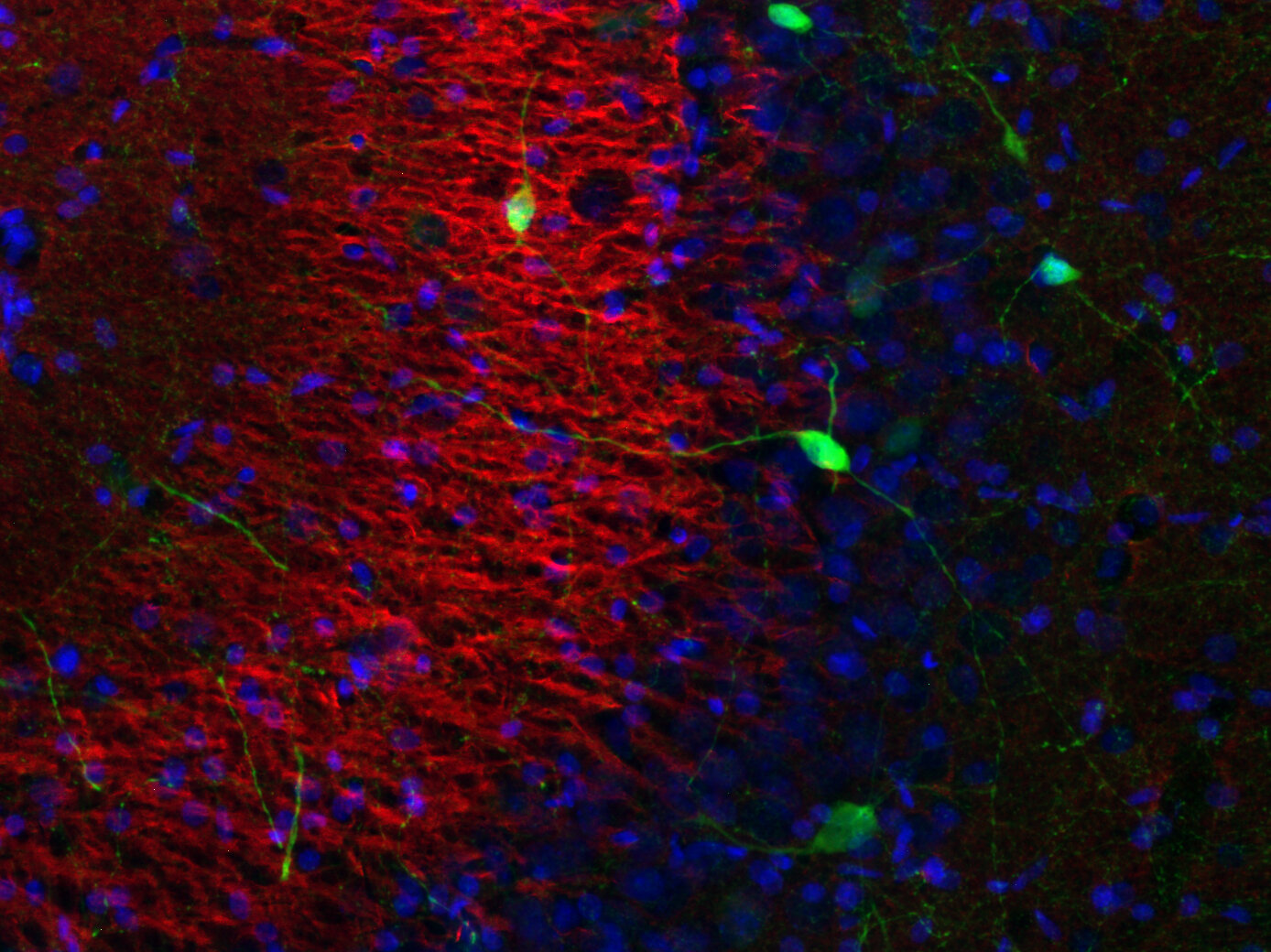
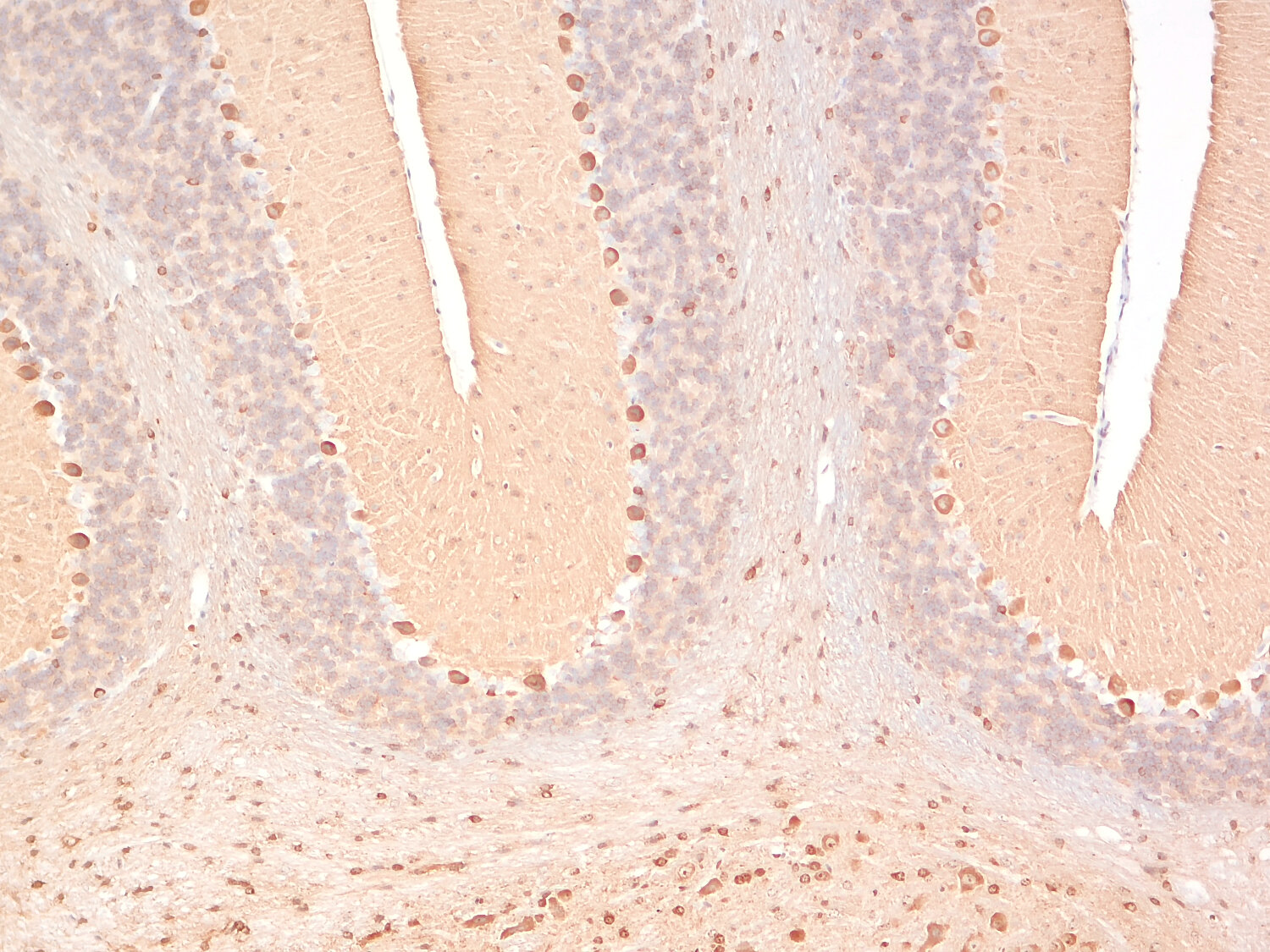
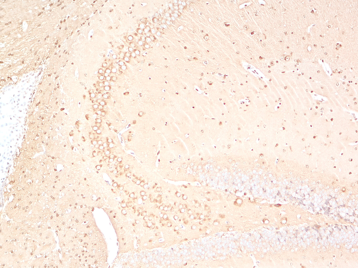
There are two major classes of heat-stable microtubule-associated proteins (MAPs): MAP2 and Tau (MAPT).
Tau is expressed in several isoforms in human brain (Tau-A, 2N4R/Tau-F, 1N4R/Tau-E, 0N4R/Tau-D, 2N3R/Tau-C, 1N3R/Tau-B, 0N3R) and rodents (Tau-A, 2N4R/Tau-F, 1N4R/Tau-E, 0N4R/Tau-D) (1). Tau helps to stabilize axonal microtubules and modulate axonal transport, with isoform diversity and phosphorylation status determining their dynamics and affinity for microtubules. Tauopathies, often associated with abnormal phosphorylation (2, 3), can be classified according to the Tau isoforms present in the pathological inclusions. For instance, Pick's disease (PiD) is characterized by tangles containing 3R-Tau isoforms (0N3R, 1N3R, and 2N3R), whereas 4R-Tau (0N4R, 1N4R, and 2N4R) accumulates in disorders like progressive supranuclear palsy (PSP) and corticobasal degeneration (CBD). In Alzheimer's disease (AD) aggregates consist of all Tau isoforms (1).
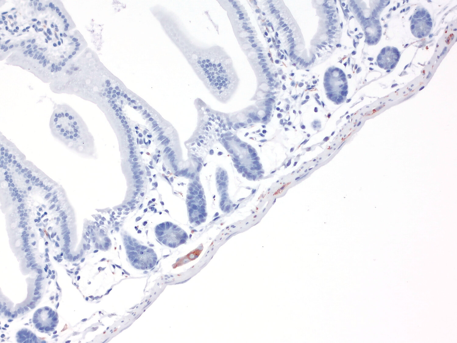
Tau is abundantly expressed in the central and peripheral nervous system. Compared to the CNS, the PNS shows a predominance of 4R Tau isoforms (0N4R, 1N4R, 2N4R), which are thought to provide stronger microtubule binding and stability needed for long peripheral axons (1, 4).
Since microtubule dynamics are central to cell division, migration, and morphology, aberrations in Tau expression have been implicated in several types of cancer (5). Notably, Tau is increasingly recognized for its role in tumor progression and resistance to cancer therapy, with glioblastoma (GBM), making Tau a potential biomarker and therapeutic target (6,7).
Certificates
ISO 9001 2015 Quality Management System and Green Lab Platinum certification level for sustaining laboratory processes.


Newsletter
Sign up for our newsletter and get the latest updates and news.