Cat. No.: 302 308
Amount: 50 µg
Price:
$420.00
|
|

|
|
| Cat. No. 302 308 |
50 µg purified recombinant IgG, lyophilized. Albumin and azide were added for stabilization. For reconstitution add 50 µl H2O to get a 1mg/ml solution in PBS. Then aliquot and store at -20°C to -80°C until use. Antibodies should be stored at +4°C when still lyophilized. Do not freeze! |
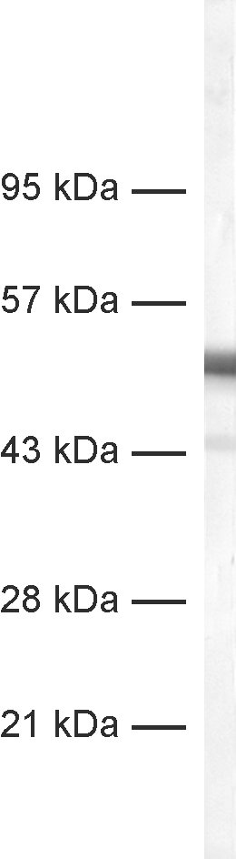
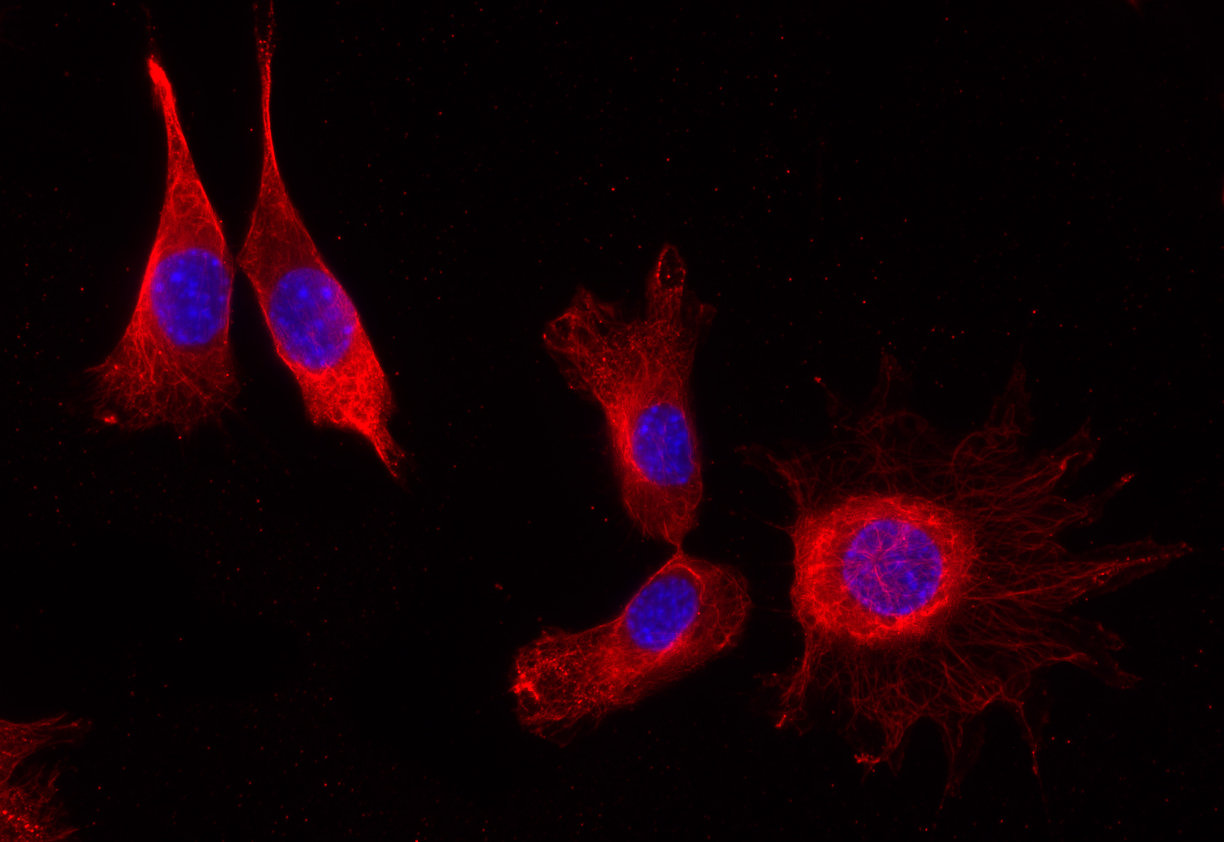
| Applications | |
| Clone | Gp3A2 |
| Subtype | IgG2 (κ light chain) |
| Immunogen | Synthetic peptide corresponding to residues near the carboxy terminus of human α-Tubulin 4A. (UniProt Id: P68366) |
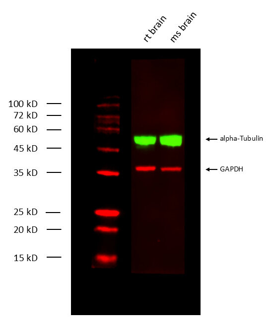
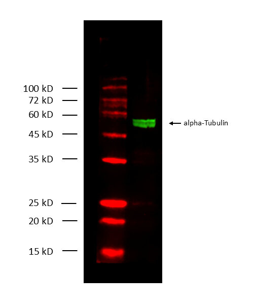
| Reactivity |
Reacts with: human (P68366), rat (Q5XIF6), mouse (P68368), vertebrates, invertebrates, yeast. Other species not tested yet. |
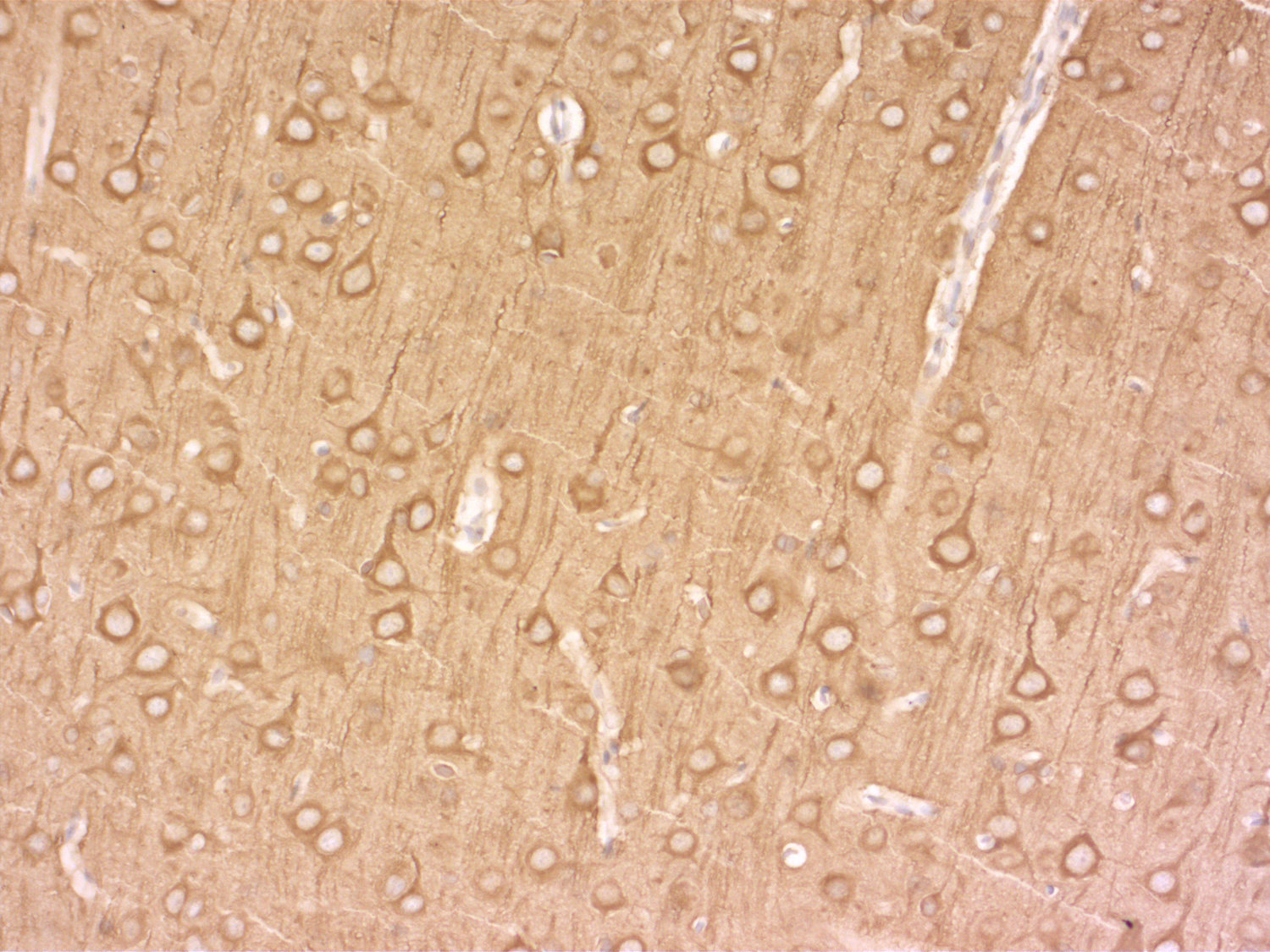
| Specificity | Specific for α-tubulin (glu- and tyr-α-tubulin) |
| Matching control protein/peptide | 302-21P |
| Remarks |
This antibody is a chimeric antibody based on the well known monoclonal mouse antibody clone 3A2. The constant regions of the heavy and light chains have been replaced by Guinea pig specific sequences. Therefore, the antibody can be used with standard anti-Guinea pig secondary reagents. The antibody has been expressed in mammalian cells. |
| Data sheet | Datasheet 302_308 |
|
|
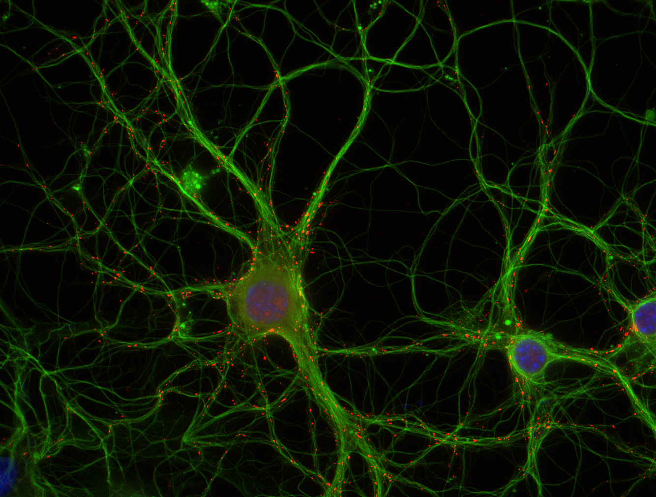
Microtubules are involved in a wide variety of intracellular events including cell division, intracellular transport and secretion, axonal transport, and maintenance of cell morphology. They are composed of tubulin, a heterodimeric protein, consisting of two polypeptides, α-tubulin and β-tubulin (1).
α Tubulin undergoes numerous post-translational modifications that include tyrosination-detyrosination and deglutamylation, phosphorylation, acetylation, polyglutamylation, and polyglycylation. In one of the major posttranslational modifications, the C-terminal tyrosine residue in α-tubulin is added or removed reversibly, producing Glu-tubulin (after detyrosination) and Tyr-tubulin (with re-added tyrosine). Early stages of cell development are often enriched in Tyr tubulin, whereas mature cells show increased Glu tubulin in stable structures. Some microtubule associated proteins (MAPs), motor proteins like kinesins, or stabilizing factors have different affinities for Glu- or Tyr-tubulin (2,3,4).
A third variant of detyrosinated α-tubulin is Δ2-tubulin which lacks the C-terminal glutamic acid. It cannot be tyrosinated by tyrosine ligase and is one of the dominant α-tubulin isoforms in neurons (5).
Certificates
ISO 9001 2015 Quality Management System and Green Lab Platinum certification level for sustaining laboratory processes.


Newsletter
Sign up for our newsletter and get the latest updates and news.