Tailor-made Antibodies
and Tools for Life Science
Home | | | | | Technical Support

GABA-A receptor alpha1 antibody extracellular - 224 208AT488

GABA-A receptors are involved in inhibitory neurotransmission
Rabbit monoclonal recombinant IgG
Cat. No.: 224 208AT488
Amount: 50 µg
Price: $470.00
Cat. No. 224 208AT488 
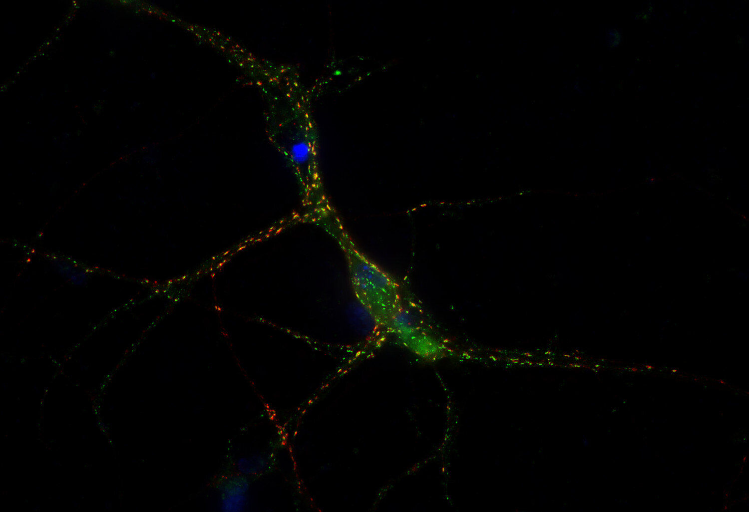
50 µg purified recombinant IgG, lyophilized, fluorescence-labeled with ATTO® 488.

For many of the fluorescence labeled antibodies conjugated to ATTO® dyes from ATTO-TEC the established fluorescence detection systems can be used.

ATTO® 488: λex 500 nm / λem 520 nm
ATTO® 550: λex 554 nm / λem 576 nm
ATTO® 565: λex 564 nm / λem 590 nm
ATTO® 594: λex 603 nm / λem 626 nm
ATTO® 647N: λex 646 nm / λem 664 nm
 

ATTO®488, ATTO®565, ATTO®647N and ATTO®594 dyes are suitable for Stimulated Emission Depletion (STED) microscopy which allows higher resolution imaging compared to confocal laser scanning microscopy.
ATTO®488, ATTO®565 and ATTO®594 are also recommended for PALM and dSTORM high resolution microscopy.

This product or portions thereof is manufactured under license from ATTO-TEC GmbH.
ATTO is a trademarks of ATTO-TEC GmbH, Siegen/Germany.
Purchase of reagents related to the AbberiorStar technology from Synaptic Systems GmbH provides a license for non-profit and in-house research use only. Any application of above mentioned technology for commercial purpose requires a separate license from ATTO-TEC GmbH.

Albumin was added for stabilization. For reconstitution add 50 µl H2O to get a 1mg/ml solution in PBS. Either add 1:1 (v/v) glycerol, then aliquot and store at -20°C until use, or store aliquots at -80°C without additives.
Reconstitute immediately upon receipt! Avoid bright light when working with the antibody to minimize photo bleeching of the fluorescent dye.
Applications
 
WB: N/A
IP: N/A
ICC: 1 : 500 (see remarks) gallery  
IHC: not tested yet
IHC-P: not tested yet

Western blot (WB); separation of proteins by PAGE and subsequent transfer to a membrane. Detection of target molecules is carried out with antibodies. Some antibodies require special sample preparation steps. For details, please refer to the “Remarks” section.

Immunoprecipitation (IP); Immunoisolation or pulldown of a target molecule using an antibody. For details and product specific hints, please refer to the ”Remarks” section.

Immunocytochemistry (ICC) on 4% PFA fixed cells. Immunoreactivity is usually revealed by fluorescence. Some antibodies require special fixation methods. For details, please refer to the “Remarks” section.

Immunohistochemistry (IHC) on 4% PFA perfusion fixed tissue with 24h PFA post fixation. Immunoreactivity is usually revealed by fluorescence or a chromogenic substrate. Some antibodies require special fixation methods or antigen retrieval steps. For details, please refer to the ”Remarks” section.

Immunohistochemistry (IHC-P) of formalin fixed, paraffin embedded (FFPE) tissue (some antibodies require special antigen retrieval steps, please refer to the ”Remarks” section). Immunoreactivity is usually revealed by fluorescence or a chromogenic substrate.

Label ATTO 488
Clone Rb53D3
Subtype IgG1 (κ light chain)
Immunogen Synthetic peptide corresponding to AA 28 to 43 from rat GABA-A receptor α1 (UniProt Id: P62813)
Reactivity Reacts with: mouse (P62812), rat (P62813).
Other species not tested yet.
Matching control protein/peptide 224-2P
Remarks

This antibody is a chimeric antibody based on the monoclonal mouse antibody clone 53D3. The constant regions of the heavy and light chains have been replaced by rabbit specific sequences.
ICC: This antibody can be used for the surface staining of living cells.

Data sheet 224_208at488.pdf
Cat. No.: 224 208AT488
Amount: 50 µg
Price: $470.00
Background

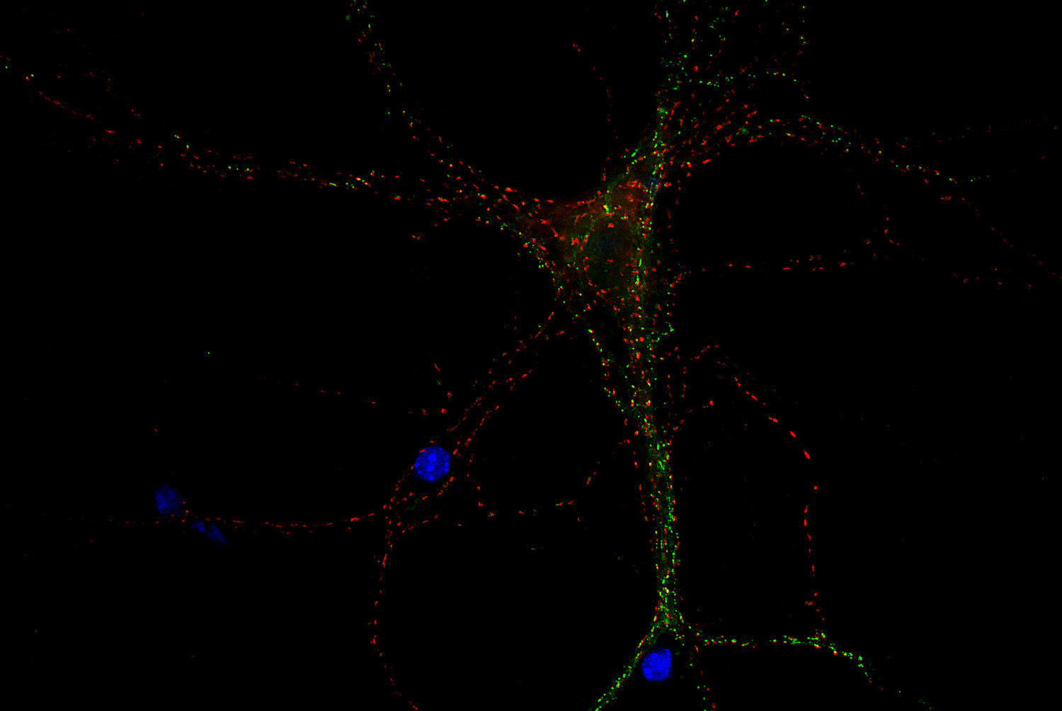
Gamma-aminobutyric acid type A (GABA-A) receptors mediate the majority of inhibitory neurotransmission in the brain. These receptor proteins are ligand gated chloride ion channels and consist of a pentameric combination of different subunits (alpha, beta, gamma, delta, epsilon and rho). The resulting heterogenous population of GABA-A receptor subtypes are expressed throughout the brain with specific cellular and subcellular expression patterns.


For more information on protein expression pattern, please refer to the overview image in our SYSY Antibodies ATLAS.