Cat. No.: 218 108
Amount: 50 µg
Price:
$420.00
|
|

|
|
| Cat. No. 218 108 |
50 µg purified recombinant IgG, lyophilized. Albumin and azide were added for stabilization. For reconstitution add 50 µl H2O to get a 1mg/ml solution in PBS. Then aliquot and store at -20°C to -80°C until use. Antibodies should be stored at +4°C when still lyophilized. Do not freeze! |
| Applications | |
| Clone | Rb2-48 |
| Subtype | IgG1 (κ light chain) |
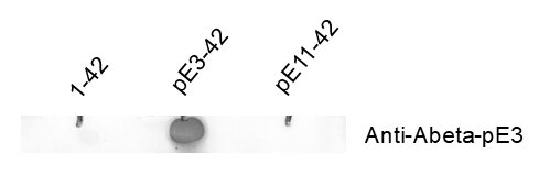
| Immunogen | Synthetic peptide corresponding to the amino terminal part of human Abeta-pE3 (UniProt Id: P05067) |
| Epitop |
AA 3 to 7 from human Abeta-pE3 (UniProt Id: P05067) |
| Reactivity |
Reacts with: human (P05067), mouse (P12023), rat (P08592), monkey. Other species not tested yet. |
| Remarks |
This antibody is a chimeric antibody based on the monoclonal mouse antibody 2-48. The constant regions of the heavy and light chains have been replaced with rabbit specific sequences. Therefore, the antibody can be used with standard anti-rabbit secondary reagents. The antibody has been expressed in mammalian cells. |
| Data sheet | Datasheet 218_108 |
|
|
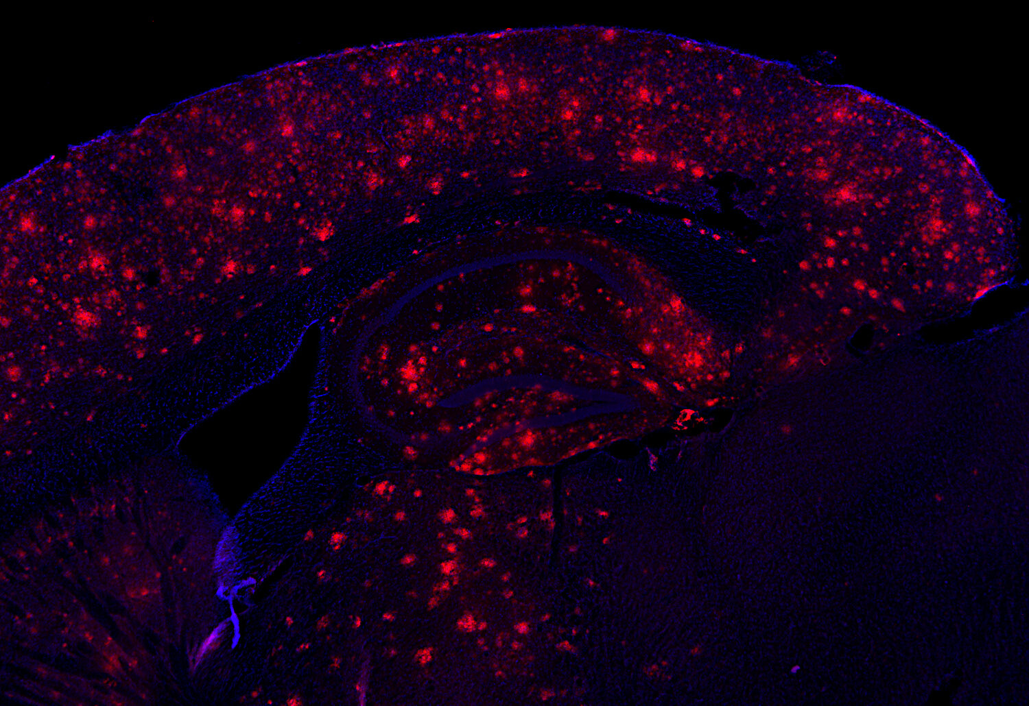
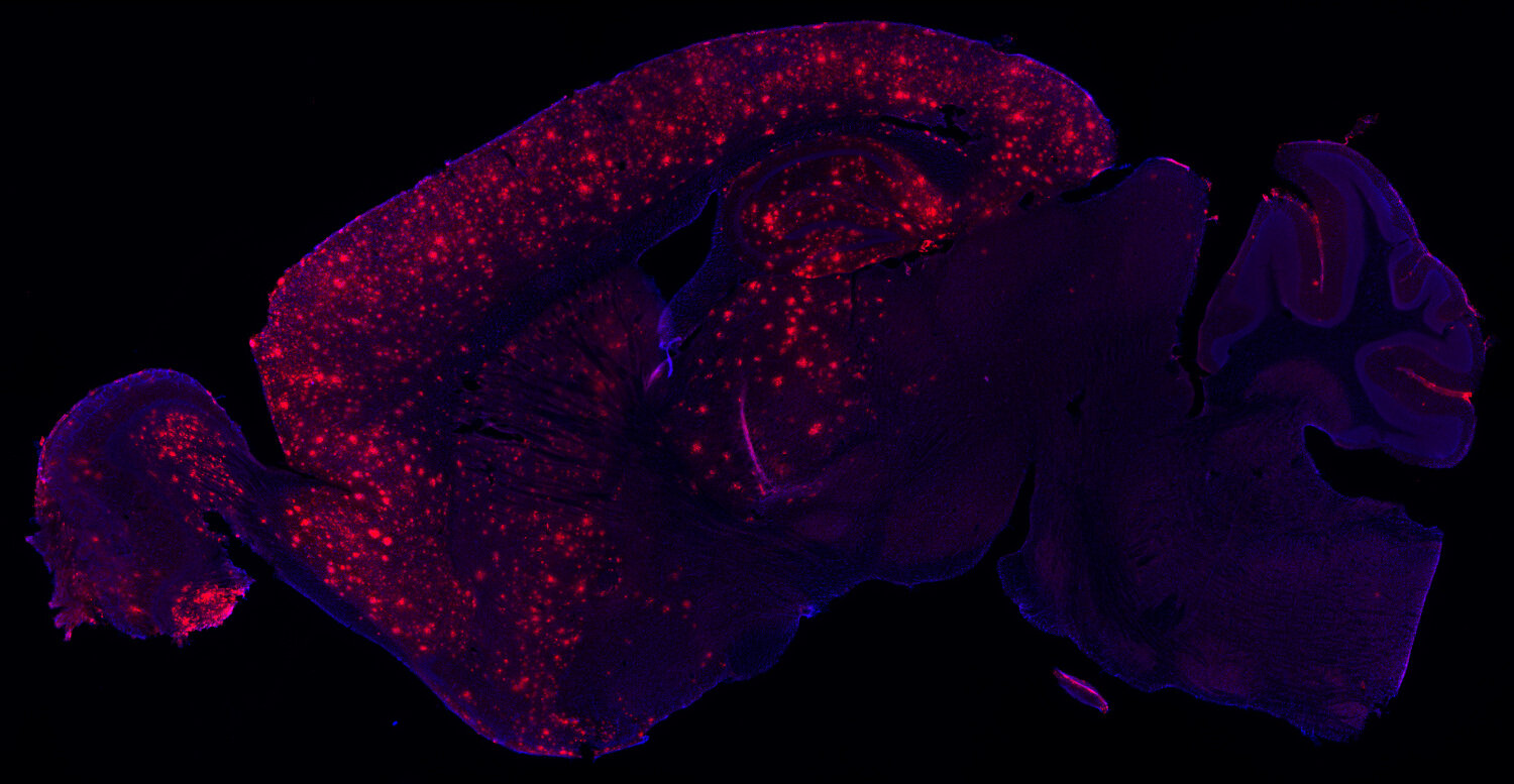
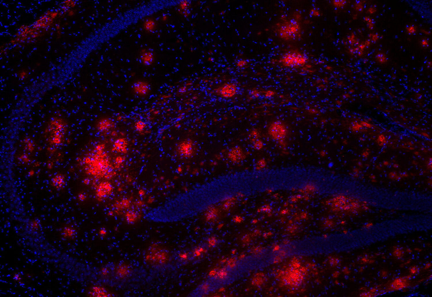
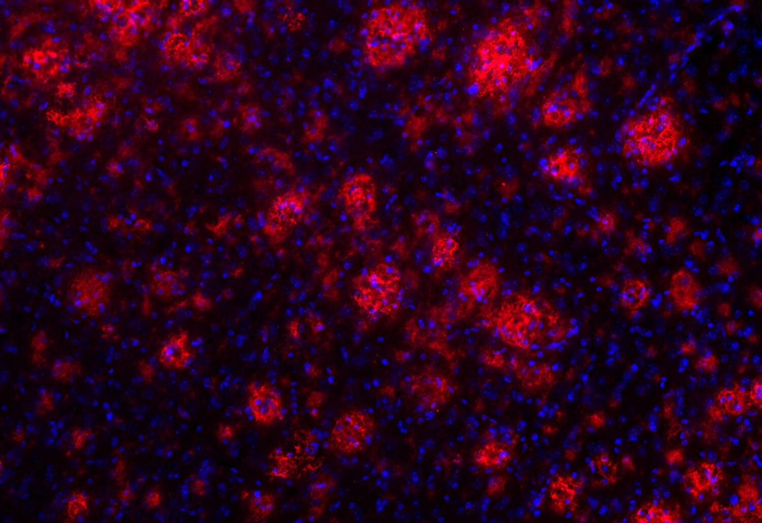
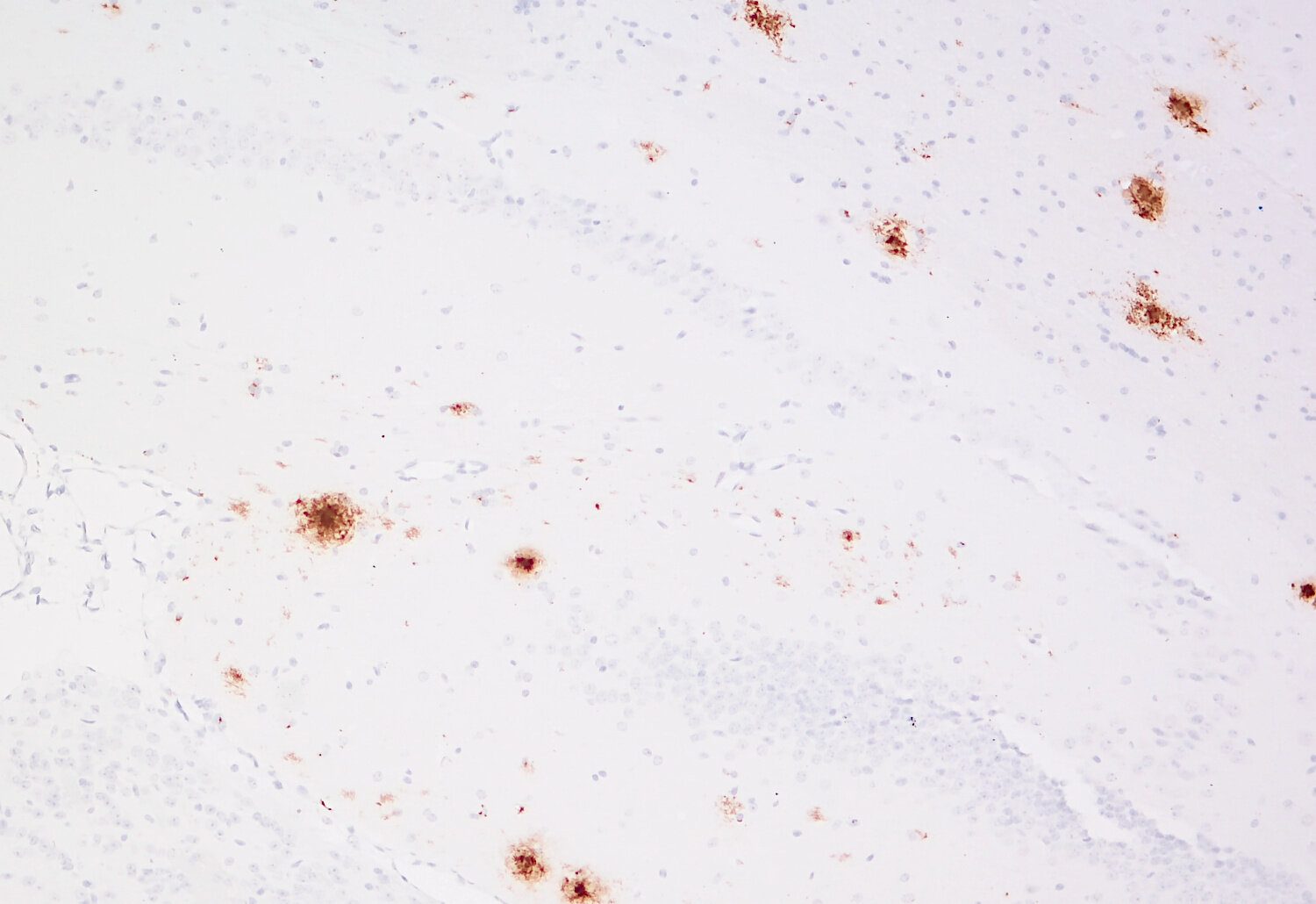
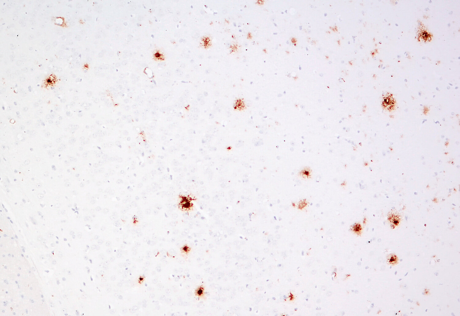
Abeta-pE3-staining of plaques (red) in a transgenic APP/PS1 Alzheimer’s disease mouse brain section
Amyloid deposits, also called plaques, of Alzheimer's patients consist of several protein components like the amyloid beta-peptides (Abeta, Aβ) 1-40/42 and additional C- and N-terminally truncated and modified fragments. Very abundant are the isoaspartate (isoAsp)-Abeta and pyroglutamyl (pGlu)-Abeta peptides. The latter are formed by cyclization of the N-terminal glutamate at position 3 or 11 catalyzed by glutaminyl cyclase (QC) resulting in very amyloidogenic and neurotoxic variants of Abeta; Abeta-pE3 and Abeta pE11.
In contrast to extracellular plaques that do not perfectly correlate with Alzheimer´s disease intraneuronal Abeta accumulation and vascular Abeta deposits have gained more and more evidence to be among the crucial factors responsible for progressive neuron loss.
Certificates
ISO 9001 2015 Quality Management System and Green Lab Platinum certification level for sustaining laboratory processes.


Newsletter
Sign up for our newsletter and get the latest updates and news.