Cat. No.: 162 106
Amount: 50 µg
Price:
$385.00
|
|

|
|
| Cat. No. 162 106 |
50 µg specific antibody, lyophilized. Affinity purified with the immunogen. Albumin and azide was added for stabilization. For reconstitution add 50 µl H2O to get a 1mg/ml solution in PBS. Then aliquot and store at -20°C to -80°C until use. Antibodies should be stored at +4°C when still lyophilized. Do not freeze! |
| Applications | |
| Immunogen | Recombinant protein corresponding to residues near the carboxy terminus of rat Shank1 (UniProt Id: Q9WV48) |
| Reactivity |
Reacts with: rat (Q9WV48), mouse (D3YZU1). Other species not tested yet. |
| Specificity | Specific for Shank 1 with weak cross-reactivity to Shank 2 and Shank 3. |
| Remarks |
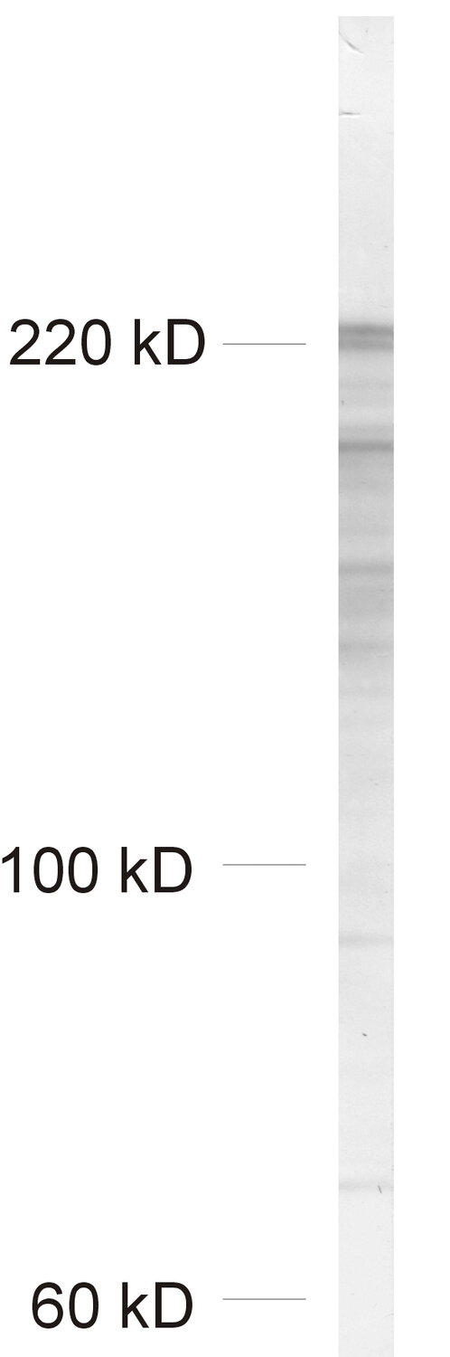
WB: Due to the large size of this protein, we recommend NuPAGE 3-8% Tris-Acetate gels for SDS-PAGE. |
| Data sheet | Datasheet 162_106 |
|
|
Shank1, 2 and 3 are major proteins of the postsynaptic density (PSD). They are composed of several protein-protein interaction domains like PDZ-, homer- and ABP1-binding domains which allow them to crosslink ionotopic and metabotropic glutamate receptor complexes with each other and to the actin-cytoskeleton.
Certificates
ISO 9001 2015 Quality Management System and Green Lab Platinum certification level for sustaining laboratory processes.


Newsletter
Sign up for our newsletter and get the latest updates and news.