Cat. No.: 106 011C3
Amount: 100 µg
Price:
$480.00
|
|

|
|
| Cat. No. 106 011C3 |
100 µg purified IgG, lyophilized, fluorescence-labeled with
Cyanine 3.
Albumin was added for stabilization. For reconstitution add 100 µl H2O to get a 1mg/ml solution in PBS. Either add 1:1 (v/v) glycerol, then aliquot and store at -20°C until use, or store aliquots at -80°C without additives. Reconstitute immediately upon receipt! Avoid bright light when working with the antibody to minimize photo bleeching of the fluorescent dye. |
| Applications | |
| Label | Sulfo-Cyanine 3 |
| Clone | 46.1 |
| Subtype | IgG1 |
| Immunogen | full-length recombinant rat Synapsin1 (UniProt Id: P09951) |
| Epitop |
AA 435 to 475 from rat Synapsin1 (UniProt Id: P09951) |
| Reactivity |
Reacts with: human (P17600), rat (P09951), mouse (O88935), mammals. Weaker signal: zebrafish, chicken, other vertebrates. Other species not tested yet. |
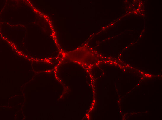
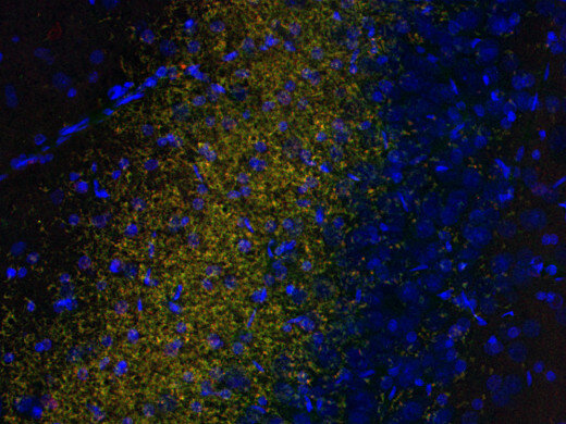
| Specificity | Specific for synapsin 1a and 1b independent of phosphorylation state. K.O. validated |
| Data sheet | Datasheet 106_011c3 |
|
|
Synapsins are neuron-specific phosphoproteins that play a fundamental role in synaptic vesicle trafficking and neurotransmitter release. They are exclusively associated with small synaptic vesicles in presynaptic terminals, with little or no expression in non-neuronal tissues including neuroendocrine cells (1–4). In mammals, three distinct genes—SYN1, SYN2, and SYN3—encode more than eight isoforms through alternative splicing. Synapsin1 is one of the most specific markers of synapses throughout both the central and peripheral nervous systems. In addition to presynaptic terminals, it is localized to sensory nerve endings and peripheral innervation of the gastrointestinal tract, including the small intestine, where it contributes to neurotransmitter release in enteric and extrinsic nerves (2,3). Two splice variants, synapsin1a and synapsin1b, interact with synaptic vesicle membranes and the cytoskeletal proteins actin and spectrin (1). Synapsin2, also expressed in the nervous system, exists in at least two splice variants, whereas synapsin3 displays a more restricted distribution, being enriched in hippocampal neurons and developing neural circuits (4).
Synapsins are major neuronal phosphoproteins and substrates of several kinases, including PKA, CaMK I, and CaMK II, with synapsin1 serving as a reference substrate for calmodulin-dependent protein kinases (1,4). Beyond their established neuronal role, recent studies have implicated synapsins in glioblastoma biology. In particular, synapsin3 has been shown to promote neuronal-like differentiation of glioblastoma stem cells by antagonizing Notch signaling, thereby reducing tumor stemness and progression (5). Moreover, glioblastoma cells can exploit synaptic communication pathways, underscoring a broader role for synaptic proteins in tumor growth and plasticity (6).
Certificates
ISO 9001 2015 Quality Management System and Green Lab Platinum certification level for sustaining laboratory processes.


Newsletter
Sign up for our newsletter and get the latest updates and news.