Cat. No.: 101 009
Amount: 50 µg
Price:
$420.00
|
|

|
|
| Cat. No. 101 009 |
50 µg purified recombinant IgY, lyophilized. Albumin and azide were added for stabilization. For reconstitution add 50 µl H2O to get a 1mg/ml solution in PBS. Then aliquot and store at -20°C to -80°C until use. Antibodies should be stored at +4°C when still lyophilized. Do not freeze! |
| Applications | |
| Clone | Ch7.2 |
| Subtype | IgY (λ light chain) |
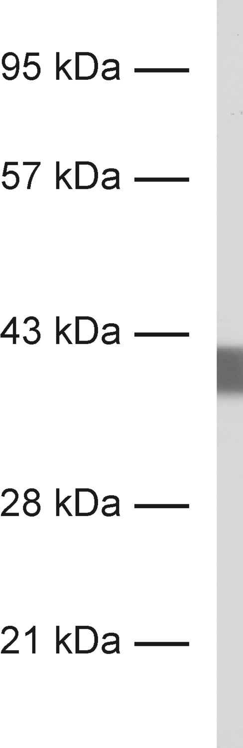
| Immunogen | Full-length recombinant rat synaptophysin (UniProt Id: P07825) |
| Epitop |
AA 219 to 307 from rat Synaptophysin1 (UniProt Id: P07825) |
| Reactivity |
Reacts with: human (P08247), rat (P07825), mouse (Q62277), other mammals. Weaker signal: zebrafish, other vertebrates. Other species not tested yet. |
| Specificity | K.O. validated |
| Remarks |
This antibody is a chimeric antibody based on the well known monoclonal mouse antibody clone 7.2. The constant regions of the heavy and light chains have been replaced by chicken specific sequences. Therefore, the antibody can be used with standard anti-chicken secondary reagents. The antibody has been expressed in mammalian cells. |
| Data sheet | Datasheet 101_009 |
|
|
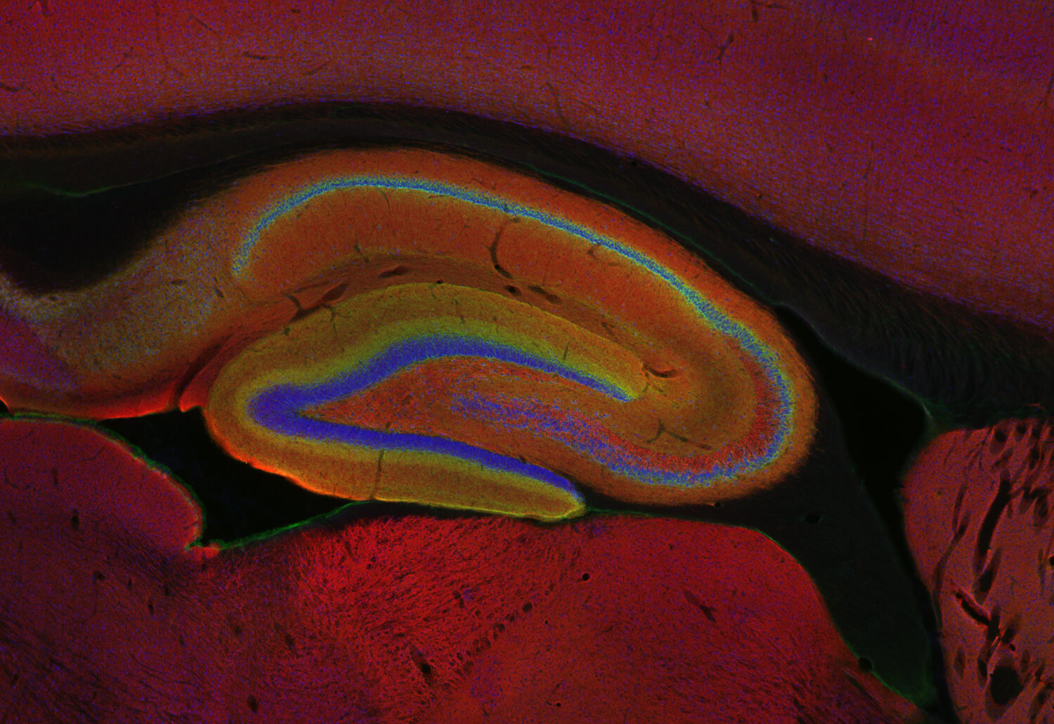
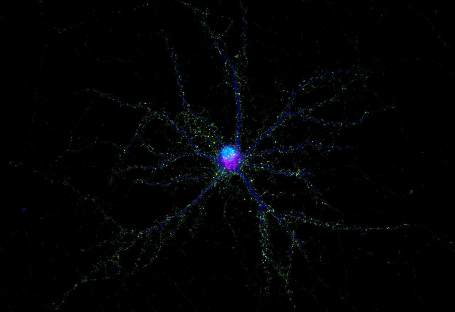
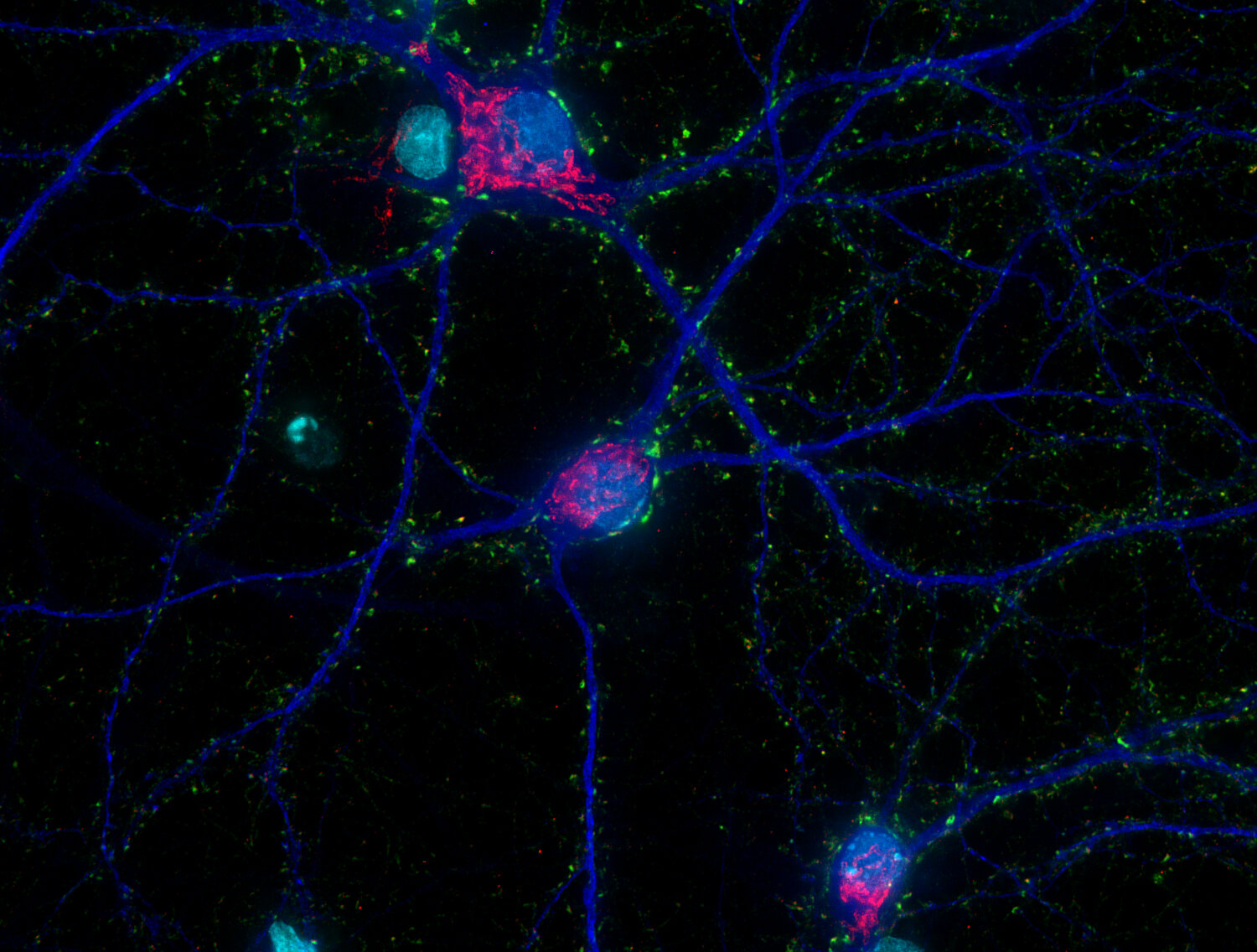
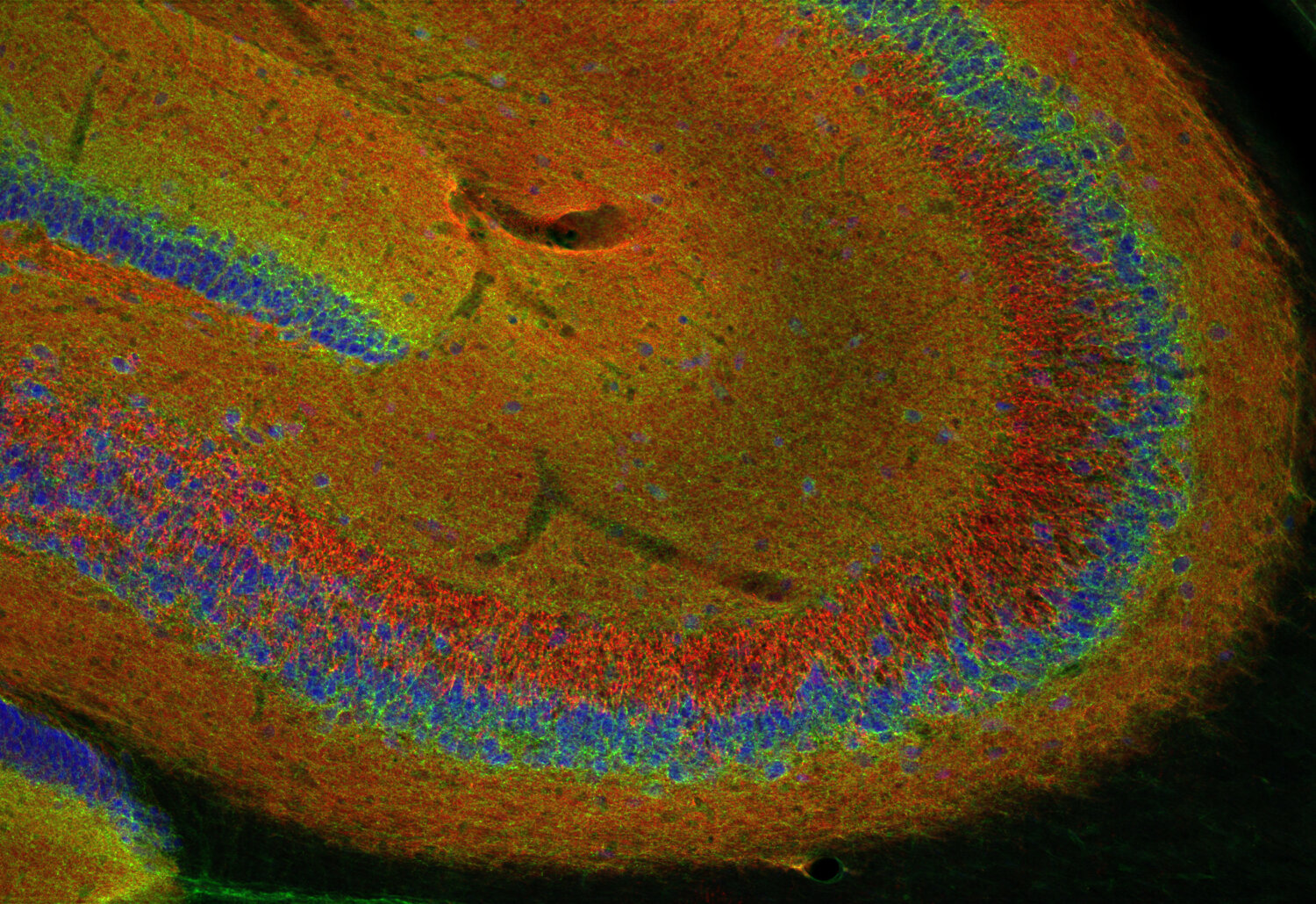
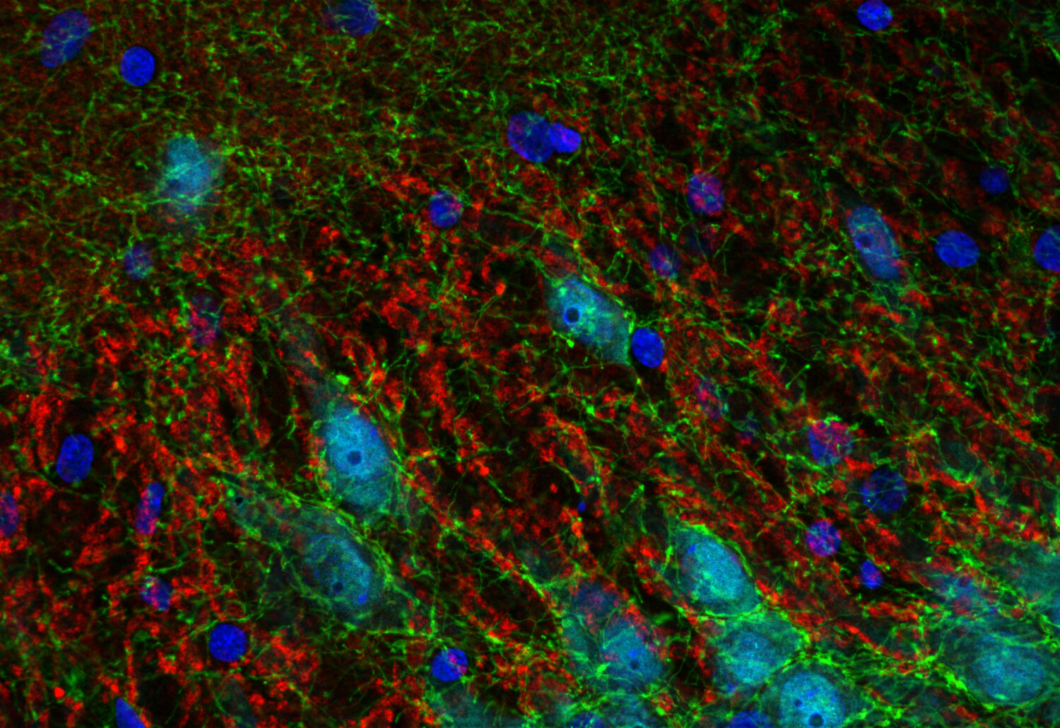
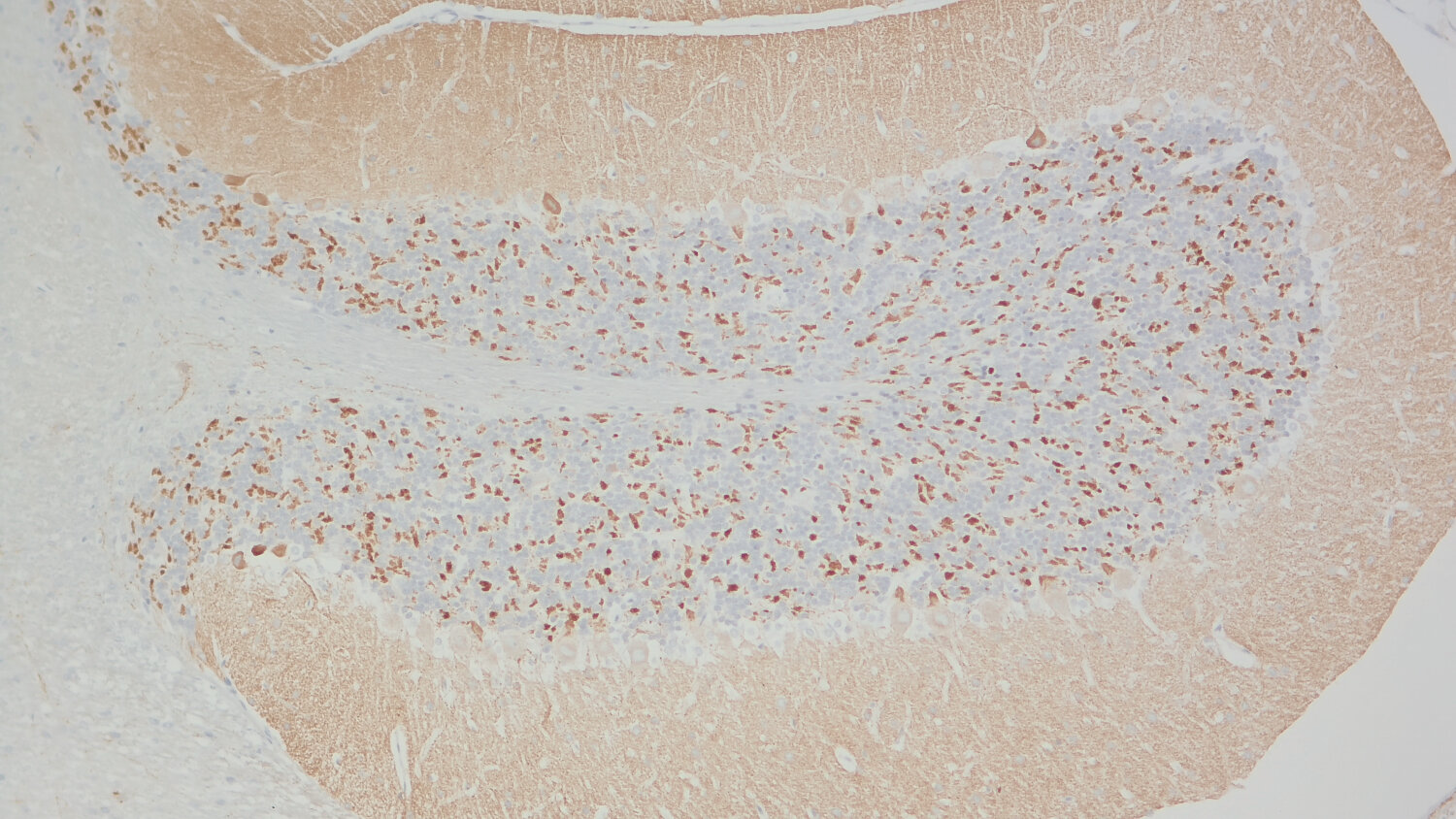
Synaptophysin1, also referred to as p38-1, is a membrane glycoprotein of synaptic vesicles that is ubiquitously expressed in all neurons and in many endocrine cells. It is currently the most widely used marker for nerve terminals and probably the best marker for the pathologist in differentiating neuroendocrine tumors.
Synaptophysin1 has four transmembrane domains with both N- and C-terminus facing the cytoplasm. It binds to synaptobrevin1 and synaptobrevin2 in detergent extracts but its function has not been elucidated completely. It forms a complex with dynamin at high Ca2+ concentration suggesting an involvement in synaptic vesicle endocytosis. As typical for synaptic vesicle proteins, synaptophysin1 represents a small protein family with two additonal members, synaptoporin (synaptophysin2) and panthophysin. Like synaptophysin1, synaptoporin is widely expressed in neurons and colocalizes with synaptophysin1 on synaptic vesicles whereas panthophysin is expressed in all tissues.
For more information on protein expression pattern, please refer to the overview image in our SYSY Antibodies ATLAS.
Certificates
ISO 9001 2015 Quality Management System and Green Lab Platinum certification level for sustaining laboratory processes.


Newsletter
Sign up for our newsletter and get the latest updates and news.